Licensing
Description
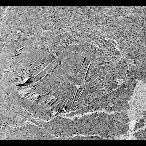
The nascent phagosome membrane has a dense coat of acidosomes docked at its cytosolic surface well before the nascent vacuole pinches off the cytopharynx. This micrograph is a quick-freeze deep-etch image of a transverse section through the posterior portion of the buccal cavity with a nascent vacuole bulging from the cytopharynx. Living cells were spun down in a centrifuge and a slurry of cells was mounted on a copper support before slam freezing the living cells against a highly polished copper surface. Some paramecia had been disrupted and this cell was feeding on the particulate organelles of the disrupted cells. Thus the nascent vacuole contains these particles amassed against its luminal surface. The cilia in the buccal cavity obviously efficiently sweep the particles against the dorsal posterior surface. TEM taken on 5/26/92 by R. Allen with Zeiss 10A operating at 80kV. Neg. 4,000X. Bar = 1µm. Published in J. Cell Sci. 106:411-422, 1993. Adapted with permission.
Technical Details
The raw negative was scanned with an Epson Perfection V750 Pro and this high resolution image is best used for quantitative analysis. Additional information available at (http://www5.pbrc.hawaii.edu/allen/).
Biological Sources
- NCBI Organism Classification
- Paramecium multimicronucleatum
- Cell Type
- cell by organism
- eukaryotic cell
- Eukaryotic Protist
- Ciliated Protist
- Cellular Component
- pre-autophagosomal structure membrane
- vesicle membrane
- oral apparatus
Biological Context
- Biological Process
- oral apparatus organization
- membrane to membrane docking
Attribution
- Name
- Richard Allen (University of Hawaii)
- Publisheds
- J Cell Sci. 1993 Sep;106 ( Pt 1):411-22.
- 8270639
- Link
- http://www5.pbrc.hawaii.edu/allen/
Grouping
This image is part of a group.Imaging
- Image Type
- transmission electron microscopy (TEM)
- illumination by electrons
- Imaging Mode
- electron density
- Parameters Imaged
- elastic scattering of electrons
- electron density
- elevation
- Source of Contrast
- differences in deposition of metal shadow
- Visualization Methods
- transmission electron microscopy (TEM)
- Processing History
- freeze_fracture/freeze_etch
- shadowing and plating
- recorded image
- film
- Data Qualifiers
- raw, unprocessed data
- suitable for spatial measurements
Sample Preparation
- Methods
- freeze_fracture/freeze_etch
- Relation To Intact Cell
- shadowing and plating
- freeze_fracture/freeze_etch
- isolated subcellular component
Dimensions
| Spatial Axis | Image Size | Pixel Size |
|---|---|---|
| X | 4000px | 2nm |
| Y | 3689px | 2nm |