Alternate header for print version


Licensing
Attribution Non-Commercial Share Alike:This image is licensed under a Creative Commons Attribution, Non-Commercial Share Alike License. View License Deed | View Legal Code
tweet  
*CIL – Cell Image Library accession number. Please use this to reference an image.

CIL:40503*  Cite 
Description

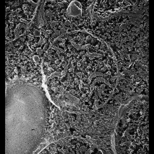
Quick-freeze deep-etch image of an unfixed and fractured mitochondrion. The tubular cristae average 50nm in diameter. Each cristae bears a helically arranged double row of particles with the particles spaced at 12 nm along this zipper-like arrangement. The cristae themselves assume a helical shape and the zipper array remains on the outside curve of the helix. In in situ fractures the 2 enclosing membranes lie close together. Crystals of unknown composition are often found in the mitochondria. TEM taken on 2/15/88 by C. Schroeder with Zeiss 10A operating at 80kV. Neg. 40,500X. Published in J. Cell Biol. 108:2233-2240, 1989. Adapted with permission. The raw negative was scanned with an Epson Perfection V750 Pro and this high resolution image is best used for quantitative analysis. Additional information available at (http://www5.pbrc.hawaii.edu/allen/).

Biological Sources
NCBI Organism Classification
Paramecium multimicronucleatum
Cell Type
cell by organism
eukaryotic cell
Eukaryotic Protist
Ciliated Protist
Cellular Component
mitochondrion
mitochondrial membrane
Biological Context
Biological Process
mitochondrion organization
Attribution
Names
Richard Allen (University of Hawaii)
C. Schroeder
Published
J Cell Biol. 1989 Jun;108(6):2233-40.
Published
2525561
Citation
Digital Object Identifier (DOI)
doi:10.7295/W9CIL40503
Archival Resource Key (ARK)
ark:/b7295/w9cil40503
Grouping This image is part of a group.
Imaging
Image Type
transmission electron microscopy (TEM)
illumination by electrons
Image Mode
electron density
Parameters Imaged
elastic scattering of electrons
electron density
elevation
Source of Contrast
differences in deposition of metal shadow
Visualization Methods
transmission electron microscopy (TEM)
Processing History
freeze_fracture/freeze_etch
shadowing and plating
recorded image
film
Data Qualifiers
raw, unprocessed data
suitable for spatial measurements
Dimensions
Spatial Axis Image Size Pixel Size
X 3870px 0.5nm
Y 4146px 0.5nm