Alternate header for print version


Licensing
Attribution Only: This image is licensed under a Creative Commons Attribution License. View License Deed | View Legal Code
tweet  
*CIL – Cell Image Library accession number. Please use this to reference an image.

CIL:40264*  Cite 
Description

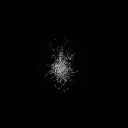
Maximum intensity projection of an optical section series of a protoplasmic astrocyte injected with Lucifer Yellow from the hippocampal area CA1 in a 1 week old male rat. For more information, see: Bushong et al. Maturation of astrocyte morphology and the establishment of astrocyte domains during postnatal hippocampal development. Int J Dev Neurosci. 2004 Apr;22(2):73-86. This image has been downsampled from the raw data image which can be accessed using the link provided to the Cell Centered Database.

Technical Details

Tissue was sectioned at a thickness of 100 µm with vibratome. Optical sections (at intervals of 0.2 µm) were gathered using a Biorad Radiance 2000 Confocal, 60X N.A. 1.4 oil-immersion objective.

Biological Sources
NCBI Organism Classification
Rattus norvegicus
Cell Type
astrocyte of the hippocampus
Cellular Component
cell body
cell projection
Attribution
Names
Eric Bushong
Link
CCDB 9
Citation
Digital Object Identifier (DOI)
doi:10.7295/W9CIL40264
Archival Resource Key (ARK)
ark:/b7295/w9cil40264
Imaging
Image Type
recorded image
Image Mode
confocal microscopy
Parameters Imaged
fluorescence emission
Visualization Methods
intracellular filling
lucifer yellow
Data Qualifiers
processed data
Sample Preparation
Methods
formaldehyde fixed tissue
Relation To Intact Cell
microtome-sectioned tissue
Dimensions
Spatial Axis Image Size Pixel Size
X 300px ——
X 235px ——