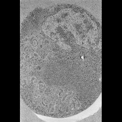
Electron micrograph of a cultured Drosophila DL1 cell infected with flock house virus, prepared by high pressure freezing followed by freeze substitution. This cell was prepared as part of an experiment to investigate different protocols for high pressure freezing. This image has been downsampled from the raw data image which can be accessed using the link provided to the Cell Centered Database.
Cell pellets were directly placed into brass planchettes that then were loaded in to the HPM 010 high pressure freezer and fast frozen. Freeze substitution: After freezing, samples (2) and (3) were placed into a Leica EM AFS Freeze substitution (FS) machine (Leica Microsystems, Bannockburn, IL) and incubated at -90 deg C for 24 hours in 0.1 percent tannic acid in acetone. Samples were washed three times with cold acetone (cooled to -90 degrees C) over 5 minutes, and placed in 1 percent OsO4 and 0.1% UA in cold acetone for 72 hours and held at -90 degrees C. After slowly warming to room temperature at 5 degrees C per hour, the specimens were rinsed in pure acetone three times (10 min. at room temperature). Infiltration and embedding in Durcupan resin was subsequently performed at room temperature. Images were acquired using a JEOL4000EX IVEM, magnification: 30000.0; accelerating voltage: 80.0 keV.
| Spatial Axis | Image Size | Pixel Size |
|---|---|---|
| X | 512px | —— |
| Y | 512px | —— |