Alternate header for print version


Licensing
Public Domain: This image is in the public domain and thus free of any copyright restrictions. However, as is the norm in scientific publishing and as a matter of courtesy, any user should credit the content provider for any public or private use of this image whenever possible. Learn more
tweet  
*CIL – Cell Image Library accession number. Please use this to reference an image.

CIL:40113*  Cite 
Description

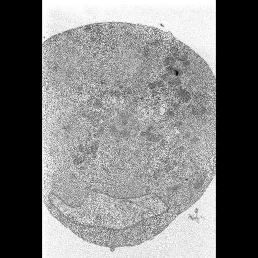
Electron micrograph of a cultured Drosophila DL1 cell infected with flock house virus, prepared using chemical fixation followed by routine embedding for electron microscopy. This specimen was prepared as a control to compare different high pressure freezing protocols. This image has been downsampled from the raw data image which can be accessed using the link provided to the Cell Centered Database.

Technical Details

Cell pellets were conventionally prepared for electron microscopy by incubation in 2% glutaraldehyde in 100 mM cacodylate approx 5 min) and then on ice for 30 minutes, followed by 1 percent osmium tetroxide in double distilled water for 1 hour and 2 percent UA in water overnight. Images were acquired using a JEOL4000EX IVEM, magnification: 30000.0; accelerating voltage: 80.0 keV.

Biological Sources
NCBI Organism Classification
Drosophila melanogaster
Cell Type
embryonic cell
Cellular Component
cell body
Attribution
Names
Mark Ellisman
Gina Sosinsky
Ying Jones
Link
CCDB 3937
Citation
Digital Object Identifier (DOI)
doi:10.7295/W9CIL40113
Archival Resource Key (ARK)
ark:/b7295/w9cil40113
Grouping This image is part of a group.
Imaging
Image Type
recorded image
Image Mode
Parameters Imaged
electron density
Data Qualifiers
processed data
Sample Preparation
Methods
osmium tetroxide fixed tissue
glutaraldehyde fixed tissue
Relation To Intact Cell
dispersed cells in vitro
Dimensions
Spatial Axis Image Size Pixel Size
X 512px ——
Y 512px ——