Alternate header for print version


Licensing
Attribution Only: This image is licensed under a Creative Commons Attribution License. View License Deed | View Legal Code
tweet  
*CIL – Cell Image Library accession number. Please use this to reference an image.

CIL:39989*  Cite 
Description

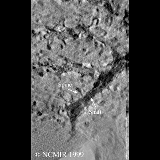
A single computed slice through tomographic volume of selectively stained Purkinje cell spiny dendrite from rat cerebellar cortex. This image has been downsampled from the raw data image which can be accessed using the link provided to the Cell Centered Database.

Technical Details

Intracellular injection with Lucifer Yellow followed by photooxidation. Images were acquired using an Hitachi 3MeV UHVEM, magnification, 4000.0, accelerating voltage, 3.0 MeV.

Biological Sources
NCBI Organism Classification
Rattus norvegicus
Cell Type
Purkinje cell
Cellular Component
dendrite
dendritic spine
Attribution
Names
Maryann Martone
Naoko Yamada
Gordun Arbuthnott
Cali Ingham
Stephen Young
Link
CCDB 3494
Citation
Digital Object Identifier (DOI)
doi:10.7295/W9CIL39989
Archival Resource Key (ARK)
ark:/b7295/w9cil39989
Imaging
Image Type
recorded image
Image Mode
Parameters Imaged
electron density
Visualization Methods
intracellular filling
lucifer yellow
photoconvertible fluorescent proteins
Data Qualifiers
processed data
Sample Preparation
Methods
osmium tetroxide fixed tissue
glutaraldehyde fixed tissue
Relation To Intact Cell
microtome-sectioned tissue