Alternate header for print version


Licensing
Public Domain: This image is in the public domain and thus free of any copyright restrictions. However, as is the norm in scientific publishing and as a matter of courtesy, any user should credit the content provider for any public or private use of this image whenever possible. Learn more
tweet  
*CIL – Cell Image Library accession number. Please use this to reference an image.

CIL:39744*  Cite 
Description

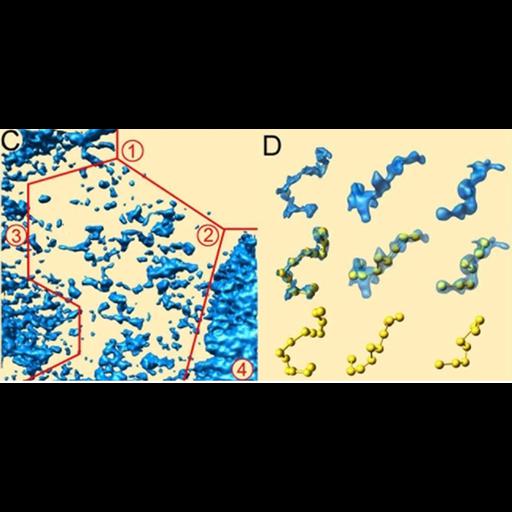
Surface rendered models of chromatin organization from tomogram derived from a TEM image of epon section of a freeze-substituted mouse retina. (C) shows the region boxed in CIL:39743 where 4 regions of chromatin compaction have been outlined. (D) shows sub-volumes interpreted as beads-on-a-string nucleosome arrays. See Fig 2C,D in: C Kizilyaprak et al. 2010. In Vivo Chromatin Organization of Mouse Rod Photoreceptors Correlates with Histone Modifications. PLOS one 5:e11039

Technical Details

See: C Kizilyaprak et al. 2010. In Vivo Chromatin Organization of Mouse Rod Photoreceptors Correlates with Histone Modifications. PLOS one 5:e11039

Biological Sources
NCBI Organism Classification
Mus musculus
Cell Type
retinal rod cell
Cellular Component
nucleus
chromatin
heterochromatin
Biological Context
Biological Process
photoreception
chromatin organization
DNA packaging
Attribution
Names
Caroline Kizilyaprak
Danièle Spehner
Didier Devys
Patrick Schultz
Published
C Kizilyaprak et al. 2010. In Vivo Chromatin Organization of Mouse Rod Photoreceptors Correlates with Histone Modifications. PLOS one 5:e11039
Pubmed
20543957
Link
PLOS one
Citation
Digital Object Identifier (DOI)
doi:10.7295/W9CIL39744
Archival Resource Key (ARK)
ark:/b7295/w9cil39744
Grouping This image is part of a group.
Imaging
Image Type
portrayed image
computer graphic
Data Qualifiers
processed data
Dimensions
Spatial Axis Image Size Pixel Size
X 899px ——
Y 447px ——