Licensing

Pd
Public Domain: This image is in the public domain and thus free of any copyright restrictions. However, as is the norm in scientific publishing and as a matter of courtesy, any user should credit the content provider for any public or private use of this image whenever possible. Learn more
Comments: Powered by Pnyxe

Description

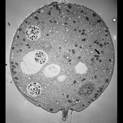
11 micrographs of views of a serially-sectioned contracted Vorticella convallaria cell that show the main features of this cell. This figure shows 5 food vacuoles and the edges of 2 more. The connection of the stalk with the cell body and the myonemes merging into the spasmoneme that passes into the contractile stalk are seen. The aboral end of the elongated macronucleus now appears. TEM taken on 4/2/71 by R. Allen with Hitachi HU11A operating at 75kV. Neg. 2,150X. The raw negative was scanned with an Epson Perfection V750 Pro and this high resolution image is best used for quantitative analysis. Additional information available at (http://www5.pbrc.hawaii.edu/allen/).

Technical Details

Standard glutaraldehyde fixation followed by osmium tetroxide, dehydrated in alcohol and embedded in an epoxy resin. Microtome sections prepared at approximately 75nm thickness. Additional information available at (http://www5.pbrc.hawaii.edu/allen/).

Biological Sources

NCBI Organism Classification
Vorticella convallaria
Cell Type
cell by organism
eukaryotic cell
Eukaryotic Protist
Ciliated Protist
Cell Line
Cambridge Culture Collection, Cambridge, England, # LB1690/1
Cellular Component
cell
food vacuole
macronucleus
contractile stalk

Biological Context

Biological Process
digestive system process
cytoplasm organization
cortical cytoskeleton organization
macronucleus organization
contractile stalk organization

Attribution

Name
Richard Allen (University of Hawaii)
Link
http://www5.pbrc.hawaii.edu/allen/

Grouping

This image is part of a group.
Data Qualifiers
raw, unprocessed data
suitable for spatial measurements

Dimensions

Spatial Axis Image Size Pixel Size
X 3635px 2.3nm
Y 4000px 2.3nm
*CIL – Cell Image Library accession number. Please use this to reference an image.