Alternate header for print version


Licensing
Attribution Non-Commercial Share Alike:This image is licensed under a Creative Commons Attribution, Non-Commercial Share Alike License. View License Deed | View Legal Code
tweet  
*CIL – Cell Image Library accession number. Please use this to reference an image.

CIL:39235*  Cite 
Description

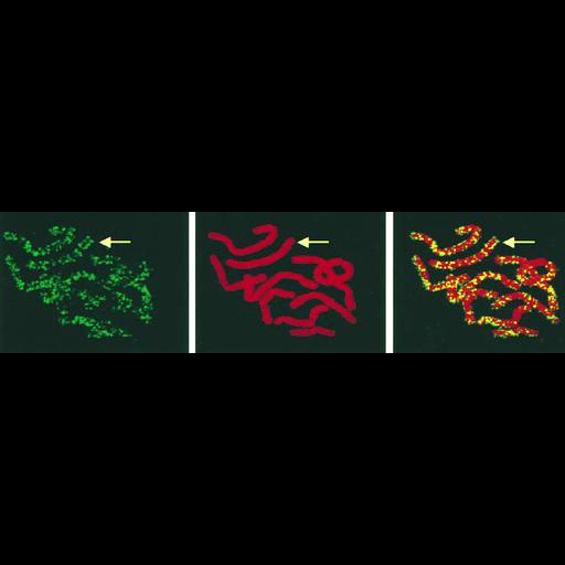
Shown is a chromosome spread of a PtK1 cell pulsed with BrdU in early S phase, then treated with colcemid to arrest cells at metaphase. Spreads were immuno-labeled with FITC-conjugated anti-BrdU antibodies and counterstained with propidium iodide (red). Replication is confined to discrete sites of replication, some of which appear to occur in bands (arrows).

Technical Details

Metaphase-arrested PtK1 Cells were swollen, pre-fixed in methanol-acetic acid, dropped onto cover glasses, incubated with H2O to promote spreading, and air dried. After hardening by a 4 hr incubation at 65C samples were fixed with 70% ethanol at -20C. Spreads were denatured with 4N HCl for 30 min at room temperature, and processed for immunofluorescence using an anti-BrdU antibody and FITC-conjugated secondary antibody. Cells were examined in a MRC-1024 confocal microscope mounted on a Nikon Optphot 2 microscope with 60x 1.4 NA objective lens. See: Fig 6 in, Ma, H et al. 1998 Spatial and temporal dynamics of DNA replication sites in mammalian cells. J Cell Biol 143:1415-1425.

Biological Sources
NCBI Organism Classification
Macropus sp. MG002
Cell Type
kidney cell
Cell Line
PtK1
Cellular Component
nuclear chromosome
Attribution
Names
Hong Ma
Jagath Samarabandu
Recandu Devdhar
Raj Acharya
Ping-chin Cheng
Chunling Meng
Ronald Berezney
Published
Ma, H et al. 1998 Spatial and temporal dynamics of DNA replication sites in mammalian cells. J Cell Biol 143:1415-1425
Pubmed
9852140
Citation
Digital Object Identifier (DOI)
doi:10.7295/W9CIL39235
Archival Resource Key (ARK)
ark:/b7295/w9cil39235
Grouping This image is part of a group.
Imaging
Image Type
photomultiplier tube (PMT)
Image Mode
confocal microscopy
Parameters Imaged
fluorescence emission
Source of Contrast
distribution of epitope
Visualization Methods
Fluorescein (FITC)
TexasRed
Processing History
montage
Data Qualifiers
processed data
Sample Preparation
Methods
ethanol fixed tissue
Relation To Intact Cell
whole mounted tissue
Dimensions
Spatial Axis Image Size Pixel Size
X 735px ——
Y 185px ——