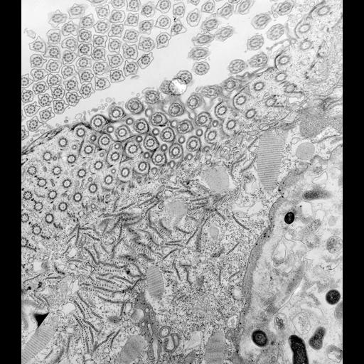
Maximum intensity project of a tomographic reconstruction of a spiny dendrite from a 4 ?m thick section throuh medium spiny neuron of mouse caudateputamen
Experiment #5 DAT KO mouse 04/22/03 Description: Photoconverted dye-filled striatal medium spiny neurons for EM Animal Info: ID# wt3 wt4 Weight: 34g 32g DOB: 9/30/02 9/30/02 Protocol 1. Perfusion (at Duke) Nembutal; 4% paraformaldehyde + 0.1% gluteraldehyde 2. Sectioned on Vibratome (at NCMIR) Thickness = 100 microns Store in 1X PBS in fridge 3. Fill cells with Lucifer yellow 4. Store slices with filled cells in 4% para in fridge 5. Wash 6x with PBS 1X (on ice) 6. When ready to begin photoconversion, turn on the chiller in confocal room. Set at ~4?C. The refrigerator unit should be set at TEMP < 45?C. Switch ON. Stage needs around 20 minutes to come to temperature. Pull unit out into hallway (to avoid increase in temperature). 6. Place slices in 2% glut/PBS on ice for 15 minutes 0.8 ml 25% gluteraldehyde 2 ml 5x PBS 6.2 ml ddH20 7. Briefly wash slices in PBS 8. Place slices in PBS/glycine for a few minutes 38 mg glycine 10 ml 1x PBS 9. Follow instructions for Photoconversion of Lucifer Yellow- filled cells 10. After photoconversion, remove DAB solution and wash slice 3x 10 minutes in generous volumes of PBS on ice. Must remove all DAB before beginning osmification. Microwaving protocol for osmication, dehydration, and embedding of photoconverted slices * Prepare Resin mix and let it sit covered and undisturbed until needed (instructions by fume hood in embedding area). * Rinse slices with a generous amount of cold 1X PBS on ice for ~ 10 min. * Turn on circulating bath (over 20?C, ~ RT): water bath (left hand side) will fill. * Insert temperature probe * Fill other T-beaker with water * Set temperature to 35?C * Open new bottle of 100% ethanol and prepare following dilutions: 90% ethanol 70% ethanol 50% ethanol * Make up osmium solution under fume hood and chill on ice * 1% osmium tetroxide in PBS on ice. 2.0 ml PBS 5X then 5.5 2x distilled H2O 2.5 ml Osmium 4% * Rinse w/ 2x distilled H2O ? 3 x 5min * Warm up microwave for 2 minutes on high * Label tubes & place in rack on ice * Fill tubes with osmium solution (w/ meniscus at 0.5) * Using glass hooks, transfer slices to tubes * Remove temperature probe & set temp above 50?C. * Put rack w. tubes in for 40 sec at full power * Change rear water load in T-beaker * Change osmium solution on ice and microwave for another 40 seconds at full power * Rinse samples for 2 minutes in distilled water on benchtop (at RT) * Insert petri bath with H2O under rack * Dehydration steps (2 x 40 seconds per step; all @ 35?C) 1st 2nd 50% EtOH 70% EtOH 90% EtOH 100% EtOH 100% Acetone * All of the dehydration steps should be carried out in microcentrifuge tubes filled with 600 ml of solution. Temperature probe should be in petri dish and set for 35. Change water in rear water load when warm to touch. * Change from water to acetone in petri bath under rack ? check acetone bath level every 3 minutes * Infiltration steps (both @ 50?C): With a 50/50 mixture of resin and acetone: 1 x 15 min 1:1 Resin:acetone * Check rear water load at 7.5 minutes Switch to 100% resin for 3 x 10 minutes: 1st 2nd 3rd 100% Resin *Periodically check rear water load * Flat embed samples between mould release slides and place in embedding oven under vacuum.
| Spatial Axis | Image Size | Pixel Size |
|---|---|---|
| X | 1024px | 0.022µm |
| Y | 1024px | 0.022µm |