Alternate header for print version


Licensing
Attribution Non-Commercial; No Derivatives:This image is licensed under a Creative Commons Attribution, Non-Commercial, No Derivatives License. View License Deed | View Legal Code
tweet  
*CIL – Cell Image Library accession number. Please use this to reference an image.

CIL:39089*  Cite 
Description

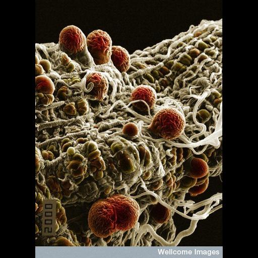
Colorized scanning electron micrograph of malaria (Plasmodium yoelii nigeriensis) oocysts ( thick-walled structure in which sporozoan zygotes develop) developing on the midgut wall of the mosquito Anopheles.

Technical Details

B0007349 Malaria parasites. Wellcome Images available under the following creative commons usage http://creativecommons.org/licenses/by-nc-nd/2.0/uk/

Biological Sources
NCBI Organism Classification
Plasmodium yoelii nigeriensis
Anopheles stephensi
Cell Type
oocyst
Cellular Component
cyst wall
Biological Context
Biological Process
sporozoan zygote development
Attribution
Names
Hilary Hurd
Link
Wellcome Images
Citation
Digital Object Identifier (DOI)
doi:10.7295/W9CIL39089
Archival Resource Key (ARK)
ark:/b7295/w9cil39089
Imaging
Image Type
recorded image
Image Mode
scanning electron microscopy (SEM)
Parameters Imaged
inelastic scattering of electrons
Source of Contrast
differences in deposition of metal shadow
Visualization Methods
visualization of contiguous regions
Processing History
colorized
Sample Preparation
Relation To Intact Cell
whole mounted tissue
Dimensions
Spatial Axis Image Size Pixel Size
X 419px ——
Y 576px ——