Alternate header for print version


Licensing
Attribution Non-Commercial; No Derivatives:This image is licensed under a Creative Commons Attribution, Non-Commercial, No Derivatives License. View License Deed | View Legal Code
tweet  
*CIL – Cell Image Library accession number. Please use this to reference an image.

CIL:39058*  Cite 
Description

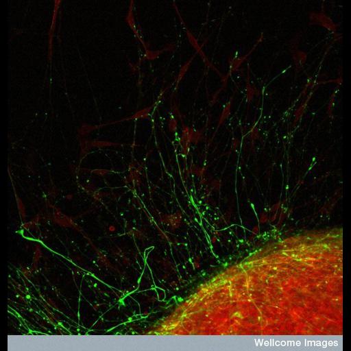
This confocal micrograph shows a dorsal root ganglion (DRG) explant. The dorsal root ganglion is a swelling on the dorsal roots of spinal nerves, which contains a cluster of cell bodies and synapses. In vertebrates the dorsal root ganglia lie outside the central nervous system. The DRG shown here has been visualized by staining with TRITC-phalloidin to highlight actin (red) and neurofilaments (green).

Technical Details

B0007573 Dorsal root ganglion. Wellcome Images available under the following creative commons usage http://creativecommons.org/licenses/by-nc-nd/2.0/uk/

Biological Context
Biological Process
neuron migration
Human Development Anatomy
dorsal root ganglion
Attribution
Names
Kevin MacKenzie
Link
Wellcome Images
OTHER
University of Aberdeen
Citation
Digital Object Identifier (DOI)
doi:10.7295/W9CIL39058
Archival Resource Key (ARK)
ark:/b7295/w9cil39058
Sample Preparation
Methods
chemically fixed tissue
permeabilized tissue
Relation To Intact Cell
explant
Dimensions
Spatial Axis Image Size Pixel Size
X 550px ——
Y 576px ——