Alternate header for print version


Licensing
Attribution Non-Commercial; No Derivatives:This image is licensed under a Creative Commons Attribution, Non-Commercial, No Derivatives License. View License Deed | View Legal Code
tweet  
*CIL – Cell Image Library accession number. Please use this to reference an image.

CIL:39012*  Cite 
Description

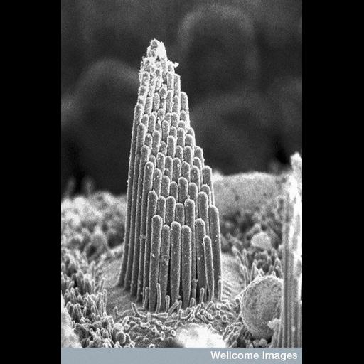
A scanning electron microscope image of the sensory hair bundle of a single hair cell from a terrapin's hearing organ in the inner ear. Vibrations made by sound cause the hairs to be moved back and forth, alternately stimulating and inhibiting the cell. When the cell is stimulated it causes nerve impulses to form in the auditory nerve, sending messages to the brain.

Technical Details

B0000112 Hair cell of inner ear. Wellcome Images available under the following creative commons usage http://creativecommons.org/licenses/by-nc-nd/2.0/uk/

Biological Sources
NCBI Organism Classification
Malaclemys terrapin
Cell Type
auditory hair cell
Cellular Component
stereocilium bundle
Attribution
Names
Dr. David Furness
Link
wellcome images
Citation
Digital Object Identifier (DOI)
doi:10.7295/W9CIL39012
Archival Resource Key (ARK)
ark:/b7295/w9cil39012
Sample Preparation
Methods
chemically fixed tissue
Relation To Intact Cell
whole mounted tissue
Dimensions
Spatial Axis Image Size Pixel Size
X 382px ——
Y 576px ——