Alternate header for print version


Licensing
Attribution Non-Commercial; No Derivatives:This image is licensed under a Creative Commons Attribution, Non-Commercial, No Derivatives License. View License Deed | View Legal Code
tweet  
*CIL – Cell Image Library accession number. Please use this to reference an image.

CIL:39006*  Cite 
Description

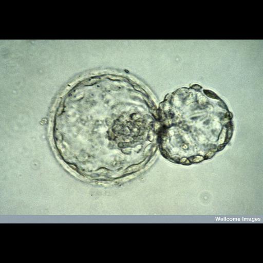
Light micrograph of a human embryo at the blastocyst stage, about six days after fertilization. The embryo is in the process of "hatching" out of the zona pellucida - the tough outer membrane - just before implanting in the wall of the uterus. The actual size of the embryo is about 0.3 mm.

Technical Details

B0000035 Human blastocyst hatching. Wellcome Images available under the following creative commons usage http://creativecommons.org/licenses/by-nc-nd/2.0/uk/

Biological Sources
NCBI Organism Classification
Homo sapiens
Cell Type
embryonic cell
Cellular Component
zona pellucida
Biological Context
Biological Process
blastocyst hatching
Human Development Anatomy
embryo
Attribution
Names
K. Hardy
Link
Wellcome Images
Citation
Digital Object Identifier (DOI)
doi:10.7295/W9CIL39006
Archival Resource Key (ARK)
ark:/b7295/w9cil39006
Imaging
Image Type
recorded image
Image Mode
modes of light microscopy
Parameters Imaged
absorption of illumination
Source of Contrast
differences in intrinsic optical density
Visualization Methods
visualization of contiguous regions
Processing History
unprocessed raw data
Sample Preparation
Relation To Intact Cell
whole mounted tissue
Dimensions
Spatial Axis Image Size Pixel Size
X 800px ——
Y 560px ——