Alternate header for print version


Licensing
Attribution Non-Commercial; No Derivatives:This image is licensed under a Creative Commons Attribution, Non-Commercial, No Derivatives License. View License Deed | View Legal Code
tweet  
*CIL – Cell Image Library accession number. Please use this to reference an image.

CIL:38991*  Cite 
Description

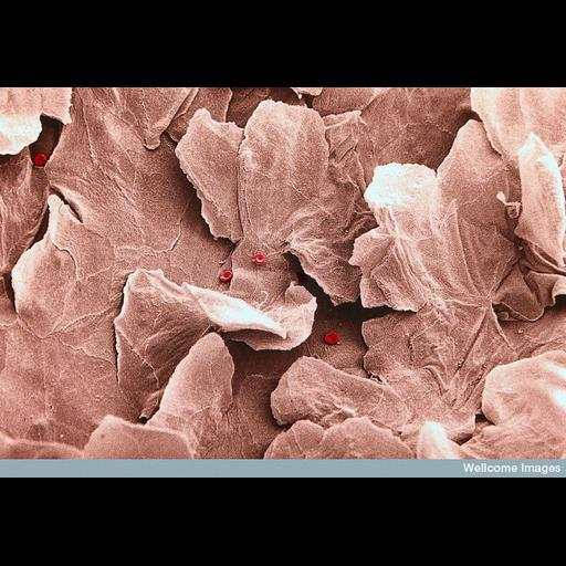
Colorized scanning electron micrograph of keratinised squamous epithelial lining of the upper vagina. The boundaries between adjacent cells are visible as raised ridges. The cells closest to the surface slough off in a similar way to skin. Five red blood cells are visible on the surface.

Technical Details

B0001965 Keratinized vaginal epithelium - coloured. Wellcome Images available under the following creative commons usage http://creativecommons.org/licenses/by-nc-nd/2.0/uk/

Biological Sources
Cell Type
keratinized squamous epithelial cell of vagina
red blood cell
Cellular Component
cell-cell junction
cell surface
Biological Context
Biological Process
epithelial cell-cell adhesion
Attribution
Names
Yorgos Nikas
Link
wellcome Images
Citation
Digital Object Identifier (DOI)
doi:10.7295/W9CIL38991
Archival Resource Key (ARK)
ark:/b7295/w9cil38991
Imaging
Image Type
recorded image
Image Mode
scanning electron microscopy (SEM)
Parameters Imaged
elastic scattering of electrons
Source of Contrast
differences in deposition of metal shadow
Visualization Methods
visualization of contiguous regions
Processing History
colorized
Sample Preparation
Methods
chemically fixed tissue
Relation To Intact Cell
whole mounted tissue
Dimensions
Spatial Axis Image Size Pixel Size
X 800px ——
Y 553px ——