Alternate header for print version


Licensing
Attribution Non-Commercial; No Derivatives:This image is licensed under a Creative Commons Attribution, Non-Commercial, No Derivatives License. View License Deed | View Legal Code
tweet  
*CIL – Cell Image Library accession number. Please use this to reference an image.

CIL:38974*  Cite 
Description

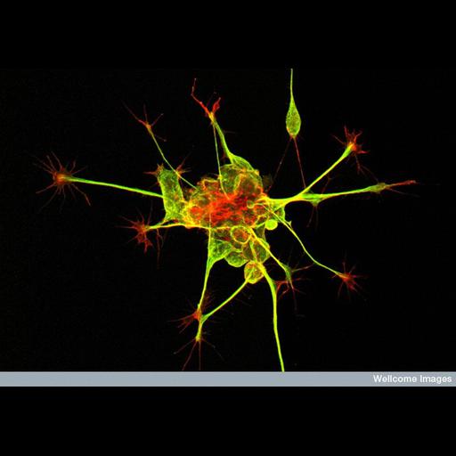
Dorsal root ganglion nerve cells stained to reveal the microtubules (green) and actin filaments (red). The axon shaft contains bundles of microtubules that give structural support and carry cargo (protein and membranes) to and from the cell body. The "hand-like" structure at the end of the axon is called the growth cone. This is very motile and reads and integrates molecular cues in the environment. It also guides the growing nerve to its target in the growing embryo.

Technical Details

B0003228 Dorsal Root Ganglion neurones in culture. Wellcome Images available under the following creative commons usage http://creativecommons.org/licenses/by-nc-nd/2.0/uk/

Biological Context
Biological Process
neuron migration
Attribution
Names
Kate Nobes
Mark Shipman
Link
Wellcome Images
Citation
Digital Object Identifier (DOI)
doi:10.7295/W9CIL38974
Archival Resource Key (ARK)
ark:/b7295/w9cil38974
Imaging
Image Type
recorded image
Image Mode
fluorescence microscopy
Parameters Imaged
fluorescence emission
Source of Contrast
distribution of epitope
Processing History
unprocessed raw data
Sample Preparation
Relation To Intact Cell
dispersed cells in vitro
Dimensions
Spatial Axis Image Size Pixel Size
X 800px ——
Y 559px ——