Alternate header for print version


Licensing
Attribution Non-Commercial; No Derivatives:This image is licensed under a Creative Commons Attribution, Non-Commercial, No Derivatives License. View License Deed | View Legal Code
tweet  
*CIL – Cell Image Library accession number. Please use this to reference an image.

CIL:38968*  Cite 
Description

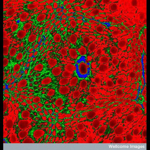
Confocal micrograph of a colonic crypt in cross section. Mucin cells (red) in the crypts produce mucous which passes into the interior of the colon through crypt lumen (the area in the centre surrounded by blue) to facilitate the movement of the faecal mass through the colon. Staining for F- actin (green) shows the pericryptal sheath. This tissue is from an endoscopic colonic biopsy for a child and shows the normal appearence of a crypt.

Technical Details

B0003962 Normal, colonic crypt with mucin cells. Wellcome Images available under the following creative commons usage http://creativecommons.org/licenses/by-nc-nd/2.0/uk/

Biological Sources
NCBI Organism Classification
Homo sapiens
Cell Type
mucin cells
Cellular Component
actin cytoskeleton
Biological Context
Biological Process
colon crypt morphology
Human Development Anatomy
colonic crypt
pericryptal sheath
colon crypt lumen
Attribution
Names
Michela Schaeppi
Link
Wellcome Images
Citation
Digital Object Identifier (DOI)
doi:10.7295/W9CIL38968
Archival Resource Key (ARK)
ark:/b7295/w9cil38968
Imaging
Image Type
recorded image
Image Mode
confocal microscopy
Parameters Imaged
fluorescence emission
Source of Contrast
distribution of epitope
Visualization Methods
primary antibody plus labeled secondary antibody
phalloidin
Processing History
unprocessed raw data
Sample Preparation
Relation To Intact Cell
sectioned tissue
Dimensions
Spatial Axis Image Size Pixel Size
X 550px ——
Y 576px ——