Alternate header for print version


Licensing
Attribution Non-Commercial; No Derivatives:This image is licensed under a Creative Commons Attribution, Non-Commercial, No Derivatives License. View License Deed | View Legal Code
tweet  
*CIL – Cell Image Library accession number. Please use this to reference an image.

CIL:38947*  Cite 
Description

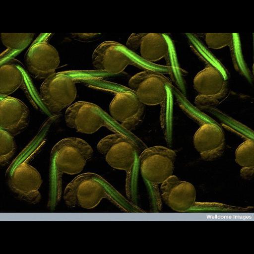
Fluorescent micrograph showing transgenic zebrafish embryos expressing green fluorescent protein in the muscle precursor cells (myotomes). The green fluorescence comes from an alpha-actin-GFP transgene acting in these living, one day old embryos.

Technical Details

B0004694 Zebrafish embryos. Wellcome Images available under the following creative commons usage http://creativecommons.org/licenses/by-nc-nd/2.0/uk/

Biological Sources
NCBI Organism Classification
Danio rerio
Cell Type
embryonic cell
muscle precursor cell
Cellular Component
actin cytoskeleton
Biological Context
Biological Process
myotome development
Zebrafish Anatomy
myotome
Attribution
Names
S. Roy
Link
Wellcome Images
Citation
Digital Object Identifier (DOI)
doi:10.7295/W9CIL38947
Archival Resource Key (ARK)
ark:/b7295/w9cil38947
Imaging
Image Type
recorded image
Image Mode
fluorescence microscopy
Parameters Imaged
fluorescence emission
Source of Contrast
distribution of a specific protein
Visualization Methods
EGFP
Processing History
unprocessed raw data
Sample Preparation
Relation To Intact Cell
whole mounted tissue
Dimensions
Spatial Axis Image Size Pixel Size
X 776px ——
Y 576px ——