Alternate header for print version


Licensing
Attribution Non-Commercial; No Derivatives:This image is licensed under a Creative Commons Attribution, Non-Commercial, No Derivatives License. View License Deed | View Legal Code
tweet  
*CIL – Cell Image Library accession number. Please use this to reference an image.

CIL:38939*  Cite 
Description

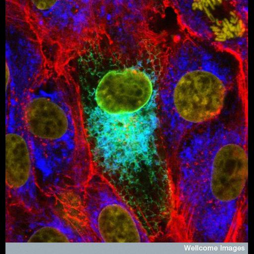
This confocal micrograph shows the mumps virus protein (turquoise) in the endoplasmic reticulum of a cultured cell. This is a region of the cell that processes proteins. This particular protein is possibly involved in determining how effectively the virus can infect people. By looking at how it works, new and more efficient vaccines could be developed. The different cells are roughly outlined in red, which stains the internal skeleton of the cells

Technical Details

B0006271 Mumps virus protein in cultured cells. Wellcome Images available under the following creative commons usage http://creativecommons.org/licenses/by-nc-nd/2.0/uk/

Biological Sources
NCBI Organism Classification
Mumps virus
Cellular Component
endoplasmic reticulum
nucleus
actin cytoskeleton
Biological Context
Biological Process
response to virus
Human Disease
mumps
Attribution
Names
Paul Duprex
Link
Wellcome Images
Citation
Digital Object Identifier (DOI)
doi:10.7295/W9CIL38939
Archival Resource Key (ARK)
ark:/b7295/w9cil38939
Imaging
Image Type
recorded image
Image Mode
confocal microscopy
Parameters Imaged
fluorescence emission
Source of Contrast
distribution of epitope
distribution of a specific protein
Processing History
unprocessed raw data
Sample Preparation
Methods
chemically fixed tissue
Relation To Intact Cell
dispersed cells in vitro
Dimensions
Spatial Axis Image Size Pixel Size
X 550px ——
Y 576px ——