Alternate header for print version


Licensing
Attribution Non-Commercial; No Derivatives:This image is licensed under a Creative Commons Attribution, Non-Commercial, No Derivatives License. View License Deed | View Legal Code
tweet  
*CIL – Cell Image Library accession number. Please use this to reference an image.

CIL:38912*  Cite 
Description

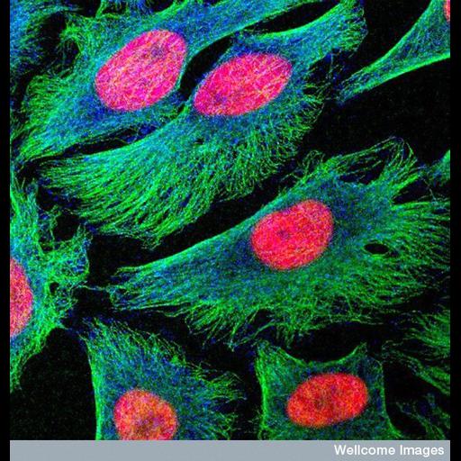
Human HeLa cancer cells in culture showing the nuclei in red and the tubulin component of the cytoskeleton in green. The blue staining is a mitotic checkpoint protein that remains in the cytoplasm until pro-metaphase when it is involved in regulating cell division. HeLa cells are derived from a human cervical cancer and can be propogated indefinitely in culture.

Technical Details

B0004985 Confocal micrograph 2003 Collection: Wellcome Images. Image available thru following license http://creativecommons.org/licenses/by-nc-nd/2.0/uk/

Biological Sources
NCBI Organism Classification
Homo sapiens
Cell Type
epithelial cell of cervix
Cell Line
HeLa
Cellular Component
nucleus
microtubule
mitotic checkpoint complex
Biological Context
Biological Process
microtubule cytoskeleton organization
Attribution
Names
Matthew Daniels
Link
Wellcome Images
Citation
Digital Object Identifier (DOI)
doi:10.7295/W9CIL38912
Archival Resource Key (ARK)
ark:/b7295/w9cil38912
Imaging
Image Type
recorded image
Image Mode
confocal microscopy
Parameters Imaged
fluorescence emission
Source of Contrast
distribution of a specific protein
Processing History
unprocessed raw data
Sample Preparation
Methods
chemically fixed tissue
Relation To Intact Cell
dispersed cells in vitro
Dimensions
Spatial Axis Image Size Pixel Size
X 550px ——
Y 576px ——