Alternate header for print version


Licensing
Attribution Non-Commercial; No Derivatives:This image is licensed under a Creative Commons Attribution, Non-Commercial, No Derivatives License. View License Deed | View Legal Code
tweet  
*CIL – Cell Image Library accession number. Please use this to reference an image.

CIL:38905*  Cite 
Description

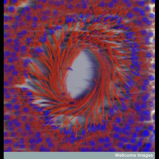
Cross section through a seminiferous tubule showing the developing sperm with their tails pointing into the lumen of the tubule. The nuclei are stained blue with DAPI and mitochondria red with mitotracker. Sperm have a large number of mitochondria to power their swim towards the egg. The image has been volume rendered using Imaris.

Technical Details

B0006849. Seminiferous tubule . Wellcome Images available under the following creative commons usage http://creativecommons.org/licenses/by-nc-nd/2.0/uk/

Biological Sources
Cell Type
sperm
Cellular Component
mitochondrion
male germ cell nucleus
Biological Context
Biological Process
spermatogenesis
Human Development Anatomy
seminiferous tubule
Attribution
Link
Wellcome Images
OTHER
MRC NIMR
Citation
Digital Object Identifier (DOI)
doi:10.7295/W9CIL38905
Archival Resource Key (ARK)
ark:/b7295/w9cil38905
Imaging
Image Type
recorded image
Image Mode
confocal microscopy
Parameters Imaged
fluorescence emission
Source of Contrast
distribution of epitope
Visualization Methods
4',6-diamidino-2-phenylindole (DAPI)
mitotracker
Processing History
volume rendering
Sample Preparation
Methods
chemically fixed tissue
Relation To Intact Cell
sectioned tissue
Dimensions
Spatial Axis Image Size Pixel Size
X 548px ——
X 576px ——