Alternate header for print version


Licensing
Attribution Non-Commercial; No Derivatives:This image is licensed under a Creative Commons Attribution, Non-Commercial, No Derivatives License. View License Deed | View Legal Code
tweet  
*CIL – Cell Image Library accession number. Please use this to reference an image.

CIL:38811*  Cite 
Description

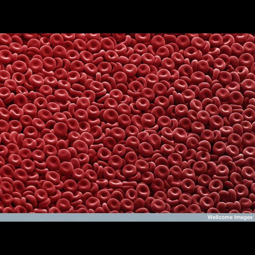
Scanning electron micrograph of red blood cells clearly showing their biconcave disc shape. Human red blood cells are typically 8 microns x 2 microns in size.

Technical Details

B0006424 2006 Collection: Wellcome Images Copyrighted work available under Creative Commons by-nc-nd 2.0 UK: England & Wales, see http://creativecommons.org/licenses/by-nc-nd/2.0/uk/

Biological Sources
Cell Type
blood cell
red blood cell
Cellular Component
cell surface
Attribution
Names
Annie Cavanagh
Link
Wellcome Images
Citation
Digital Object Identifier (DOI)
doi:10.7295/W9CIL38811
Archival Resource Key (ARK)
ark:/b7295/w9cil38811
Imaging
Image Type
recorded image
Image Mode
scanning electron microscopy (SEM)
Parameters Imaged
elastic scattering of electrons
Source of Contrast
differences in deposition of metal shadow
Visualization Methods
visualization of contiguous regions
Processing History
colorized
Sample Preparation
Methods
dispersed cells in vitro
Relation To Intact Cell
whole mounted tissue
Dimensions
Spatial Axis Image Size Pixel Size
X 780px ——
Y 576px ——