Alternate header for print version


Licensing
Attribution Non-Commercial; No Derivatives:This image is licensed under a Creative Commons Attribution, Non-Commercial, No Derivatives License. View License Deed | View Legal Code
tweet  
*CIL – Cell Image Library accession number. Please use this to reference an image.

CIL:38810*  Cite 
Description

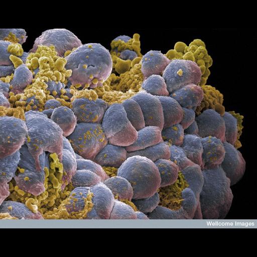
Scanning electron micrograph of a cluster of breast cancer cells showing visual evidence of programmed cell death (apoptosis) in yellow. Each cell is 15 micrometers across.

Technical Details

Image B0006422. 2006 Collection: Wellcome Images Copyrighted work available under Creative Commons by-nc-nd 2.0 UK: England & Wales, see http://creativecommons.org/licenses/by-nc-nd/2.0/uk/

Biological Sources
Cell Type
breast cancer cell
Cellular Component
cell surface
Biological Context
Biological Process
apoptotic process
Human Disease
breast carcinoma
Attribution
Names
Annie Cavanagh
Link
Wellcome Images
Citation
Digital Object Identifier (DOI)
doi:10.7295/W9CIL38810
Archival Resource Key (ARK)
ark:/b7295/w9cil38810
Imaging
Image Type
recorded image
Image Mode
scanning electron microscopy (SEM)
Parameters Imaged
elastic scattering of electrons
Source of Contrast
differences in amount of elastic light scattering
Processing History
colorized
Sample Preparation
Methods
fixed tissue
Relation To Intact Cell
whole mounted tissue
Dimensions
Spatial Axis Image Size Pixel Size
X 740px ——
Y 576px ——