Alternate header for print version


Licensing
Attribution Non-Commercial:This image is licensed under a Creative Commons Attribution, Non-Commercial License. View License Deed | View Legal Code
tweet  
*CIL – Cell Image Library accession number. Please use this to reference an image.

CIL:37289*  Cite 
Description

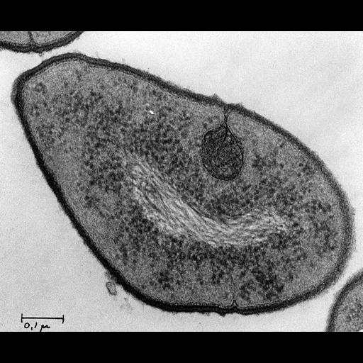
A transmission electron micrograph of a diplococcus (renamed Streptococcus in 1974) pneumoniae bacteria. A striking feature of these bacteria is an intra-cytoplasmic membrane system which appears to be an extension of septa in dividing bacteria. Image is related to Figure 3 in J. Cell. Biol. 22:453-467, 1964. Image made available by James D. Jamieson and the Department of Cell Biology, Yale University School of Medicine.

Technical Details

Original 3.25 in. x 4 in. lantern slides were scanned at 600dpi. Original magnificaiton X10,000.

Biological Sources
NCBI Organism Classification
Streptococcus pneumoniae
Cell Type
bacteria
Cellular Component
nuclear fibrils
chondroid
ribosome
Attribution
Names
James D. Jamieson
Published
J. Cell. Biol. 22:453-467, 1964.
Published
14203390
Link
George E. Palade EM Slide Collection
Citation
Digital Object Identifier (DOI)
doi:10.7295/W9CIL37289
Archival Resource Key (ARK)
ark:/b7295/w9cil37289
Grouping This image is part of a group.
Imaging
Image Type
recorded image
Image Mode
transmission electron microscopy (TEM)
Parameters Imaged
electron density
Source of Contrast
differences in adsorption or binding of stain
Visualization Methods
lead salt
Sample Preparation
Relation To Intact Cell
sectioned tissue
Dimensions
Spatial Axis Image Size Pixel Size
X 6000px ——
Y 4976px ——