Alternate header for print version


Licensing
Public Domain: This image is in the public domain and thus free of any copyright restrictions. However, as is the norm in scientific publishing and as a matter of courtesy, any user should credit the content provider for any public or private use of this image whenever possible. Learn more
tweet  
*CIL – Cell Image Library accession number. Please use this to reference an image.

CIL:36793*  Cite 
Description

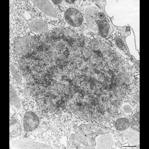
The micronucleus of Paramecium caudatum is distinctive from other closely related Paramecium such as P. multimicronucleatum and P. tetraurela. P. caudatum has only one micronucleus of a type in which the chromatin is spread equally throughout the nucleus as opposed to P. multimicronucleatum which has four very small micronuclei of the vesicular type and P. aurelia which has two micronuclei of the vesicular type where the chromatin forms a central electron opaque mass in the center of the nucleus. The micronucleus in this micrograph contains many microtubules so it may be in early division. The nuclear envelope has many nuclear pores. TEM taken on 3/23/73 by R. Allen with Hitachi HU11A operating at 75kV. Neg. 13,250X. Bar = 0.5µm.

Technical Details

Standard glutaraldehyde fixation followed by osmium tetroxide, dehydrated in alcohol and embedded in an epoxy resin. Microtome sections prepared at approximately 75nm thickness. The negative was printed to paper and the image was scanned to Photoshop. This digitized image is available for qualitative analysis. There is a high resolution version of this image in the library (CIL:39204) which is available for quantitative analysis. Additional information available at (http://www5.pbrc.hawaii.edu/allen/).

Biological Sources
NCBI Organism Classification
Paramecium caudatum
Cell Type
cell by organism
eukaryotic cell
Eukaryotic Protist
Ciliated Protist
Cellular Component
micronucleus
microtubule associated complex
nuclear pore
Biological Context
Biological Process
micronucleus organization
micronucleus division
Attribution
Names
Richard Allen (University of Hawaii)
Citation
Digital Object Identifier (DOI)
doi:10.7295/W9CIL36793
Archival Resource Key (ARK)
ark:/b7295/w9cil36793
Grouping This image is part of a group.
Imaging
Image Type
transmission electron microscopy (TEM)
illumination by electrons
Image Mode
detection of electrons
film
Parameters Imaged
electron density
Source of Contrast
stain with broad specificity
Visualization Methods
stain with broad specificity
uranyl salt
lead salt
osmium tetroxide
Processing History
recorded image
film
Print from negative scanned for Photoshop.
Data Qualifiers
processed data
Dimensions
Spatial Axis Image Size Pixel Size
X 3074px ——
Y 3508px ——