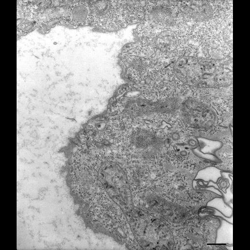
The tubules arising from late DV-III expand at their distal ends into rounded shapes. These expanded ends contain a glycocalyx like the secondary lysosomes and large IMPs also like secondary lysosomes. TEM taken on 8/7/80 by R. Allen with Hitachi HU11A TEM. Neg. 12,250X. Bar = 0.5µm. Part published in J. Cell Biol. 99:1955-1959, 1984. Adapted with permission.
Standard glutaraldehyde fixation followed by osmium tetroxide, dehydrated in alcohol and embedded in an epoxy resin. Microtome sections prepared at approximately 75nm thickness. The negative was printed to paper and the image was scanned to Photoshop. This image is suitable for qualitative analysis. A high resolution version of this image in the library (CIL:40565) is available for quantitative analysis. Additional information available at (http://www5.pbrc.hawaii.edu/allen/).