Alternate header for print version


Licensing
Public Domain: This image is in the public domain and thus free of any copyright restrictions. However, as is the norm in scientific publishing and as a matter of courtesy, any user should credit the content provider for any public or private use of this image whenever possible. Learn more
tweet  
*CIL – Cell Image Library accession number. Please use this to reference an image.

CIL:36752*  Cite 
Description

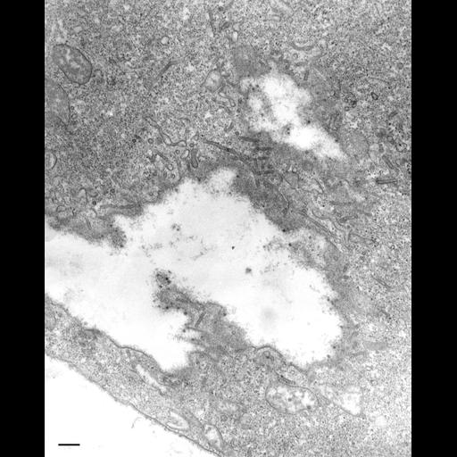
Following digestion, that only occurs in the phagolysosome (DV-III), the membrane of these DVs undergoes extensive tubulation. These tubules are ~45nm in diameter and arise from the cytosolic surface of the DV-III in clumps of 3 to 6 tubules per clump. The tubules contain a glycocalyx-like lining in their lumens which leaves only a 10nm diameter opening in the center of the tubule (see Allen and Fok, J. Cell Biol. 99:1955-1959, 1984). TEM taken on 4/13/79 by R. Allen with Hitachi HU11A operating at 75kV. Neg. 14,750X. Bar = 0.25µm.

Technical Details

Standard glutaraldehyde fixation followed by osmium tetroxide, dehydrated in alcohol and embedded in an epoxy resin. Microtome sections prepared at approximately 75nm thickness. The negative was printed to paper and the image was scanned to Photoshop. This digitized image is available for qualitative analysis. Additional information available at (http://www5.pbrc.hawaii.edu/allen/).

Biological Sources
NCBI Organism Classification
Paramecium multimicronucleatum
Cell Type
cell by organism
eukaryotic cell
Eukaryotic Protist
Ciliated Protist
Cellular Component
phagolysosome
Biological Context
Biological Process
digestive system process
Attribution
Names
Richard Allen (University of Hawaii)
Citation
Digital Object Identifier (DOI)
doi:10.7295/W9CIL36752
Archival Resource Key (ARK)
ark:/b7295/w9cil36752
Imaging
Image Type
transmission electron microscopy (TEM)
illumination by electrons
Image Mode
detection of electrons
film
Parameters Imaged
electron density
Source of Contrast
stain with broad specificity
Visualization Methods
stain with broad specificity
osmium tetroxide
uranyl salt
lead salt
Processing History
recorded image
film
Print from negative scanned for Photoshop.
Data Qualifiers
processed data
Dimensions
Spatial Axis Image Size Pixel Size
X 2107px ——
Y 2622px ——