Alternate header for print version


Licensing
Attribution Non-Commercial Share Alike:This image is licensed under a Creative Commons Attribution, Non-Commercial Share Alike License. View License Deed | View Legal Code
tweet  
*CIL – Cell Image Library accession number. Please use this to reference an image.

CIL:36745*  Cite 
Description

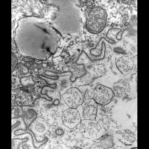
The entire membrane of the phagolysosome does appear to have been changed into that similar to the lysosome membrane which bears a thick glycocalyx (polysaccharide coat) and no longer appears to be like the phagoacidosome which does not have a thick glycocalyx. As no patches of acidosome-like membrane remain it can be assumed that the acidosome-like membrane has been removed. Fibrous material lines the cytosolic side of the early DV-III similar to that found at docking sites between the lysosomes and the DV-II. TEM taken on 3/28/80 by R. Allen with Hitachi HU11A operating at 75kV. Neg. 18,000X. Bar = 0.2µm.

Technical Details

Standard glutaraldehyde fixation followed by osmium tetroxide, dehydrated in alcohol and embedded in an epoxy resin. Microtome sections prepared at approximately 75nm thickness. The negative was printed to paper and the image was scanned to Photoshop. This digitized image is available for qualitative analysis. Additional information available at (http://www5.pbrc.hawaii.edu/allen/).

Biological Sources
NCBI Organism Classification
Paramecium multimicronucleatum
Cell Type
cell by organism
eukaryotic cell
Eukaryotic Protist
Ciliated Protist
Cellular Component
phagolysosome
Biological Context
Biological Process
digestive system process
Attribution
Names
Richard Allen (University of Hawaii)
Citation
Digital Object Identifier (DOI)
doi:10.7295/W9CIL36745
Archival Resource Key (ARK)
ark:/b7295/w9cil36745
Imaging
Image Type
transmission electron microscopy (TEM)
illumination by electrons
Image Mode
detection of electrons
film
Parameters Imaged
electron density
Source of Contrast
stain with broad specificity
Visualization Methods
stain with broad specificity
osmium tetroxide
uranyl salt
lead salt
Processing History
recorded image
film
Print from negative scanned for Photoshop.
Data Qualifiers
processed data
Dimensions
Spatial Axis Image Size Pixel Size
X 2112px ——
Y 2496px ——