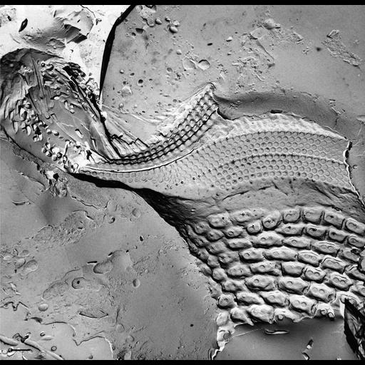
The oral region, illustrated here in a freeze-fracture image that passed through its left side, opens at the mid-ventral part of the cell. It consists of a funnel shaped vestibulum covered with regular ciliature arising from normal somatic surface depressions. The vestibulum opens into the tube-like buccal cavity through an oral overture. On the anterior dorsal surface and left side of the buccal cavity are the three complex ciliary membranelles: the ventral peniculus, dorsal peniculus and quadrulus. Each membranelle is composed of four rows of closely packed cilium/basal body complexes. Posterior to the vestibulum the floor of the buccal cavity is supported on the cytosolic side by a filamentous reticulum which also covers the nonciliated ribbed wall on the right side of the buccal cavity. The posterior dorsal half of the buccal cavity opens through the cytostome into the cytopharynx and the developing food vacuole. Since the structures of this oral complex in P. multimicronucleatum are the same as those of P. caudatum. TEM taken on 6/2/73 by D. Leaffer with Hitachi HU11A operating at 75kV. Neg. 3,500X. Bar = 1µm. Published in J. Cell Biol. 63:904-922, 1974. Adapted with permission.
| Spatial Axis | Image Size | Pixel Size |
|---|---|---|
| X | 3179px | —— |
| Y | 3096px | —— |