Alternate header for print version


Licensing
Public Domain: This image is in the public domain and thus free of any copyright restrictions. However, as is the norm in scientific publishing and as a matter of courtesy, any user should credit the content provider for any public or private use of this image whenever possible. Learn more
tweet  
*CIL – Cell Image Library accession number. Please use this to reference an image.

CIL:36287*  Cite 
Description

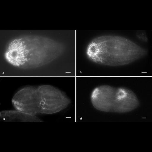
Immunofluorescent pictures of Tetrahymena labeled with an antibody to the V-ATPase of Dictyostelium discoideum provided by Dr. T. L. Steck, University of Chicago. a. This polyclonal antibody was the same as that used in Fok et al., J. Cell Sci. 108:3163-3170, 1995, to label the decorated tubules of Paramecium. The decorated tubules form a striated cap over the posterior end of the cell but leave the CV pore and the CV itself unlabeled. b. During division the cap is reduced in size in the opisthe while in figure c a new field of decorated tubules appears near the equatorial region of the dividing cell near the posterior end of the proter. d. Both decorated-tubule regions grow in size and in intensity as division proceeds. Fluorescent micrographs taken by M. Aihara on 12/6/91 and 1/3/92. Unpublished results. Bars = 20µm. The negative was printed to paper and the image was scanned to Photoshop. This digitized image is available for qualitative analysis. Additional information available at (http://www5.pbrc.hawaii.edu/allen/).

Biological Sources
NCBI Organism Classification
Tetrahymena pyriformis
Cell Type
cell by organism
eukaryotic cell
Eukaryotic Protist
Ciliated Protist
Cellular Component
contractile vacuole
vacuolar proton-transporting V-type ATPase, V0 domain
Biological Context
Biological Process
contractile vacuole organization
Attribution
Names
Richard Allen (University of Hawaii)
M. Aihara
Citation
Digital Object Identifier (DOI)
doi:10.7295/W9CIL36287
Archival Resource Key (ARK)
ark:/b7295/w9cil36287
Imaging
Image Type
fluorescence microscopy
Image Mode
fluorescence microscopy
Parameters Imaged
compartmentalization of stain or label
Source of Contrast
fluorescence microscopy
Visualization Methods
macromolecular probe
Processing History
recorded image
Print scanned for Photoshop.
Data Qualifiers
processed data
Sample Preparation
Methods
glutaraldehyde fixed tissue
Relation To Intact Cell
whole mounted tissue
Dimensions
Spatial Axis Image Size Pixel Size
X 3612px ——
Y 2190px ——