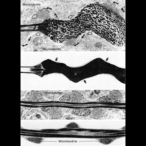
Figure 426 (A-D) from Chapter 16 (Cytoplasmic matrix and cytoskeleton) of 'The Cell, 2nd Ed.' by Don W. Fawcett M.D. Stages of differentiation of the finch spermatozoon. A.)Early in spermatogenesis, a large bundle of micortubules winds a helical course around the condensign nucleus as the head is beginning to take on its mature corkscrew-shaped form. B.) Although the microtubules disappear after the shape develops, shallow depressions at the surface of the head (arrows) mark their former location. C.)The microtubule bundle continues posteriorly and winds around the midpiece forming a double helix with the continus strand of fused mitochondria. D.)The mitochondrial helix of the mature sperm retains the same pitch as the microtubule bundle after the latter has disappeared. Image from Fawcett et al., Dev. Biol. 26:220-251 (1971). A PDF copy of the accompanying chapter is available on the ASCB’s BioEDUCATE website.
| Spatial Axis | Image Size | Pixel Size |
|---|---|---|
| X | 901px | —— |
| Y | 1254px | —— |