Alternate header for print version


Licensing
Attribution Non-Commercial; No Derivatives:This image is licensed under a Creative Commons Attribution, Non-Commercial, No Derivatives License. View License Deed | View Legal Code
tweet  
*CIL – Cell Image Library accession number. Please use this to reference an image.

CIL:36049*  Cite 
Description

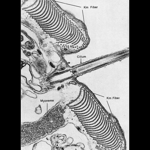
Figure 414 from Chapter 16 (Cytoplasmic matrix and cytoskeleton) of 'The Cell, 2nd Ed.' by Don W. Fawcett M.D. Transverse section of a portion of the cortex of a partially extended Stentor coerulus shows a cilium flanked by two microtubule arrays in cross section. The microtubules are interconnected by short bridges to form ribbons that are attached to basal bodies of neighboring cilia. During extension, the overlapping ribbons of stiff microtubules slide with respect to on another, decreasing their degree of overlap and causing the organism to elongate. Underlying myonemes contract to shorten the organism. From Huang and Pitelka, J. Cell Biol. 57:704-728, 1973 (PMID:4633444). A PDF copy of the accompanying chapter is available on the ASCB’s BioEDUCATE website.

Biological Sources
NCBI Organism Classification
Stentor coeruleus
Cellular Component
microtubule
cilium
Biological Context
Biological Process
microtubule sliding
cilium movement involved in cell motility
Attribution
Names
Don W. Fawcett
B. Huang; D.R. Pitelka
Published
ISBN 0721635849
Link
http://www.ascb.org/bioeducate/FawcettTheCell...
Citation
Digital Object Identifier (DOI)
doi:10.7295/W9CIL36049
Archival Resource Key (ARK)
ark:/b7295/w9cil36049
Grouping This image is part of a group.
Imaging
Image Type
transmission electron microscopy (TEM)
Image Mode
illumination by electrons
detection of electrons
Parameters Imaged
electron density
Source of Contrast
stain with broad specificity
Visualization Methods
electron-dense stain
Data Qualifiers
processed data
Dimensions
Spatial Axis Image Size Pixel Size
X 882px ——
Y 1218px ——