Alternate header for print version


Licensing
Attribution Non-Commercial; No Derivatives:This image is licensed under a Creative Commons Attribution, Non-Commercial, No Derivatives License. View License Deed | View Legal Code
tweet  
*CIL – Cell Image Library accession number. Please use this to reference an image.

CIL:36044*  Cite 
Description

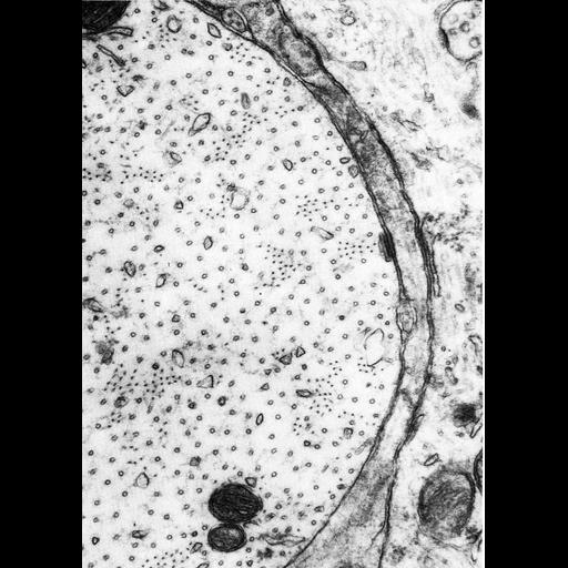
Figure 407 from Chapter 16 (Cytoplasmic matrix and cytoskeleton) of 'The Cell, 2nd Ed.' by Don W. Fawcett M.D. Proximal dendrite from a neuron of the anterior horn of the spinal cord. In this micrograph, longitudinally-oriented microtubules are observed in cross-section, and distributed uniformly throughout the cytoplasm. Loose aggregations of smaller neurofilaments (10 nm) are also present. Micrograph by Raymond Wuerker. A PDF copy of the accompanying chapter is available on the ASCB’s BioEDUCATE website.

Biological Sources
NCBI Organism Classification
Vertebrata
Cell Type
CNS neuron (sensu Vertebrata)
Cellular Component
microtubule
dendrite cytoplasm
neurofilament
Biological Context
Biological Process
microtubule cytoskeleton organization
Attribution
Names
Don W. Fawcett
Raymond Wuerker
Published
ISBN 0721635849
Link
http://www.ascb.org/bioeducate/FawcettTheCell...
Citation
Digital Object Identifier (DOI)
doi:10.7295/W9CIL36044
Archival Resource Key (ARK)
ark:/b7295/w9cil36044
Grouping This image is part of a group.
Imaging
Image Type
transmission electron microscopy (TEM)
Image Mode
illumination by electrons
detection of electrons
Parameters Imaged
electron density
Source of Contrast
stain with broad specificity
Visualization Methods
electron-dense stain
Data Qualifiers
processed data
Dimensions
Spatial Axis Image Size Pixel Size
X 898px ——
Y 1266px ——