Licensing
Attribution Only: This image is licensed under a Creative Commons Attribution License. View License Deed | View Legal Code
Comments:
Powered by Pnyxe
Description
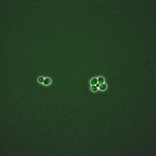
A time lapse experiment of Saccharomyces cerevisiae expressing GFP-tagged MCM1. MCM1 is a transcription factor involved in cell-type-specific transcription and pheromone response; plays a central role in the formation of both repressor and activator complexes These phase, gfp images are part of an image group that ranges from CIL:35880-35899. Note that there are additional groups showing time series of other cell cycle regulation proteins by the same authors in the Library.
Technical Details
Time-lapse images were collected on a DeltaVision system (Olympus IX71) with 60x/1.42 NA objective at 5 minute intervals. Please see the microscopy section in the referenced manuscript for details for image analysis.
Biological Sources
- NCBI Organism Classification
- Saccharomyces cerevisiae
- (Baker's Yeast, Brewer's Yeast)
- Cellular Component
- MCM1 protein
- nuclear chromatin
- nucleus
Biological Context
- Biological Process
- arginine metabolic process
- cell cycle
- regulation of transcription, DNA-dependent
- transcription, DNA-dependent
- DNA-dependent DNA replication initiation
- regulation of transcription from RNA polymerase II promoter
- Molecular Function
- DNA binding
- DNA bending activity
- DNA replication origin binding
- sequence-specific DNA binding transcription factor activity
Attribution
- Names
- David Ball
- Jean Peccoud
- Published
- PLoS One. 2011;6(10):e26272. Epub 2011 Oct 26.
- Pubmed
- 22046265
Grouping
This image is representative of a group of similar images.Imaging
- Image Type
- recorded image
- Imaging Mode
- fluorescence microscopy
- phase contrast microscopy
- Parameters Imaged
- fluorescence emission
- elastic scattering of photons
- Source of Contrast
- distribution of a specific protein
- differences in amount of elastic light scattering
- Visualization Methods
- EGFP
- visualization of contiguous regions
Sample Preparation
- Methods
- living tissue
- Relation To Intact Cell
- dispersed cells in vitro
Dimensions
| Spatial Axis | Image Size | Pixel Size |
|---|---|---|
| X | 512px | 213.4nm |
| Y | 512px | 213.4μm |
| Time | 300 sec | 61 |
*CIL – Cell Image Library accession number. Please use this to reference an image.