Alternate header for print version


Licensing
Public Domain: This image is in the public domain and thus free of any copyright restrictions. However, as is the norm in scientific publishing and as a matter of courtesy, any user should credit the content provider for any public or private use of this image whenever possible. Learn more
tweet  
*CIL – Cell Image Library accession number. Please use this to reference an image.

CIL:35610*  Cite 
Description

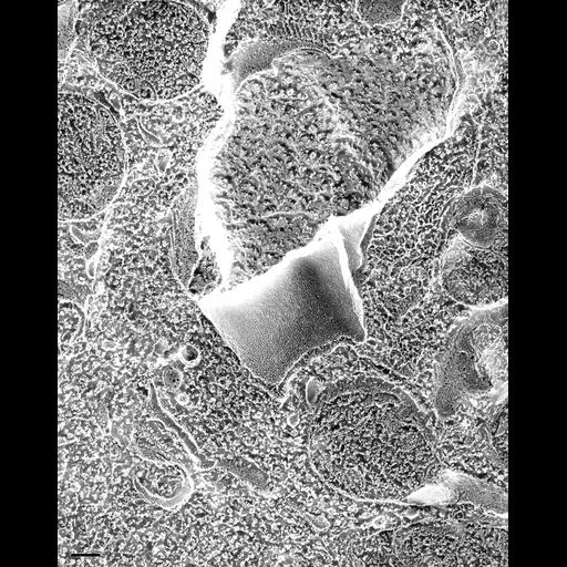
Quick-freeze deep-etch of an ampulla. The P-fracture face has numerous IMPs. The microtubular ribbon now forms two bands next to this ampulla. The ribbons will split into additional bands as they pass on to and around the collecting canal. TEM taken on 1/14/95 by R. Allen with Zeiss 10A operating at 80kV. Neg. 19,800X. Bar = 0.2µm. A print of the negative was scanned and processed in Photoshop. This image is best used for qualitative analysis. Additional information available at (http://www5.pbrc.hawaii.edu/allen/).

Biological Sources
NCBI Organism Classification
Paramecium multimicronucleatum
Cell Type
cell by organism
eukaryotic cell
Eukaryotic Protist
Ciliated Protist
Cellular Component
contractile vacuole
cytoplasm
contractile vacuolar membrane
microtubule
microtubule associated complex
Attribution
Names
Richard Allen (University of Hawaii)
Citation
Digital Object Identifier (DOI)
doi:10.7295/W9CIL35610
Archival Resource Key (ARK)
ark:/b7295/w9cil35610
Imaging
Image Type
transmission electron microscopy (TEM)
illumination by electrons
Image Mode
electron density
Parameters Imaged
elastic scattering of electrons
electron density
elevation
Source of Contrast
differences in deposition of metal shadow
Visualization Methods
transmission electron microscopy (TEM)
Processing History
freeze_fracture/freeze_etch
shadowing and plating
recorded image
film
Print of negative scanned for Photoshop
Data Qualifiers
processed data
Dimensions
Spatial Axis Image Size Pixel Size
X 2045px ——
Y 2574px ——