Alternate header for print version


Licensing
Public Domain: This image is in the public domain and thus free of any copyright restrictions. However, as is the norm in scientific publishing and as a matter of courtesy, any user should credit the content provider for any public or private use of this image whenever possible. Learn more
tweet  
*CIL – Cell Image Library accession number. Please use this to reference an image.

CIL:34846*  Cite 
Description

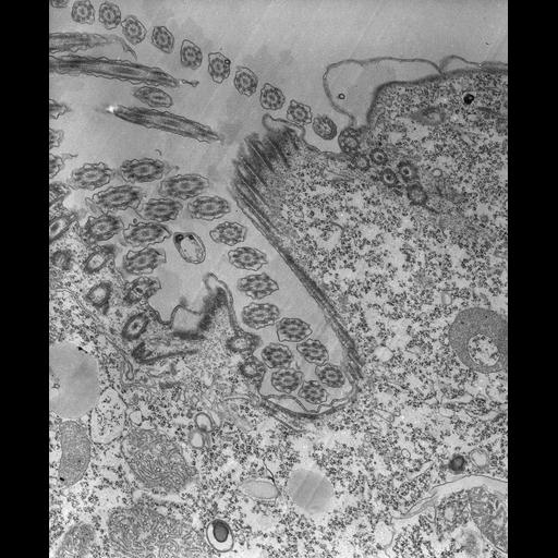
A high resolution tangential section through the buccal cavity showing the close spacing of the cilia in the undulating membrane and 3, presumably nonciliated basal bodies, in the inside row. Accessory microtubular ribbons arise from the basal bodies of the inner row and extend upward toward the oral ribs. Microtubular ribbons in the oral ribs are sectioned obliquely and alveolar segments lie between the ribbons. Portions of two membranelles are seen in the buccal cavity. TEM taken on 8/15/67 by R. Allen with Philips 200 operating at 60kV. Neg. 12,400X. The raw film was scanned with an Epson Perfection V750 Pro. This image is best used for quantitative analysis.

Technical Details

Standard glutaraldehyde fixation followed by osmium tetroxide, dehydrated in alcohol and embedded in an epoxy resin. Microtome sections prepared at approximately 75nm thickness. Additional information available at (http://www5.pbrc.hawaii.edu/allen/).

Biological Sources
NCBI Organism Classification
Tetrahymena pyriformis
Cell Type
cell by organism
eukaryotic cell
Eukaryotic Protist
Ciliated Protist
Cellular Component
oral apparatus
Biological Context
Biological Process
oral apparatus organization
Attribution
Names
Richard Allen (University of Hawaii)
Citation
Digital Object Identifier (DOI)
doi:10.7295/W9CIL34846
Archival Resource Key (ARK)
ark:/b7295/w9cil34846
Grouping This image is part of a group.
Imaging
Image Type
transmission electron microscopy (TEM)
illumination by electrons
Image Mode
detection of electrons
film
Parameters Imaged
electron density
Source of Contrast
stain with broad specificity
Visualization Methods
stain with broad specificity
osmium tetroxide
uranyl salt
lead salt
Processing History
recorded image
film
unprocessed raw data
Data Qualifiers
raw, unprocessed data
suitable for spatial measurements
Dimensions
Spatial Axis Image Size Pixel Size
X 5058px 1.2nm
Y 6150px 1.2nm