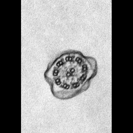
A cross section of cilium through the cilium-basal body complex extending from the cilium to the transition zone between the cilium and basal body and ending at the proximal tip of the basal body. In cross section the 9 + 2 axoneme of the cilium stands out. The plasma membrane covers the axoneme and is continuous with the plasma membrane covering the cell body. Each doublet tilts inward to the tangent of the axonemal cylinder. Two arms, an inner arm and outer arm, extend from the complete microtubule of each doublet. These arms have ATPase activity and are called dyneins. They cause one doublet to slide against the adjacent doublet. Other protein links, like the spokes, in the axoneme are elastic and induce the characteristic bending of the cilium and prevent excess sliding as the doublets slide past each other. TEM taken on 7/15/67 by R. Allen with RCA EMU3F operating at 50kV. Neg. 19,200X. Adapted with permission. The raw film was scanned with an Epson Perfection V750 Pro. This image is best used for quantitative analysis.
Standard glutaraldehyde fixation followed by osmium tetroxide, dehydrated in alcohol and embedded in an epoxy resin. Microtome sections prepared at approximately 75nm thickness. Additional information available at (http://www5.pbrc.hawaii.edu/allen/).
| Spatial Axis | Image Size | Pixel Size |
|---|---|---|
| X | 604px | 0.78nm |
| Y | 886px | 0.78nm |