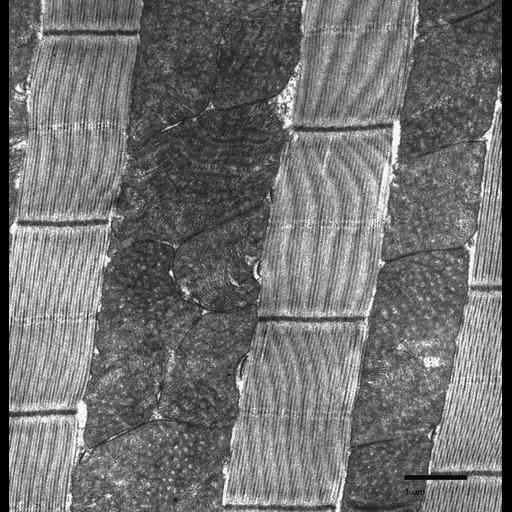
Drosophila melanogaster thoracic indirect flight muscle from a four week-old wild-type animal. A longitudinal section of myocyte is displayed. Sections of parallel myofibrils are visible between densely packed mitochondria.
Tissue was dissected using a freeze-fracture technique, fixed in 2.5% glutaraldehyde in 0.1 M sodium cacadylate buffer, pH 7.4, post-fixed in 2% osmium tetroxide solution, stained in 1% uranyl acetate, and embeddeded in EMbed 812 resin. Sections were post-stained with 2% uranyl acetate and stained with Reynolds’s Lead Citrate. Images were obtained using transmission electron microscopy techniques, employing a Phillips CM-100 transmission electron microscope. Images were taken on Kodak EM 4489 film, scanned with a Microtek ScanMaker i900, and post-processed using Adobe Photoshop CS4. This image was taken at a magnification of 11500x.
| Spatial Axis | Image Size | Pixel Size |
|---|---|---|
| X | 1766px | 42.33µm |
| Y | 1832px | 42.33µm |