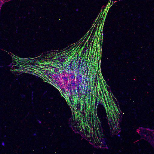
To study the molecular mechanism by which nonmuscle myosin II (MII) regulates protrusion and adhesion dynamics in migrating cells, NIH3T3 cells were transfected with myc-tagged myosin light chain (MLC, green) and co-stained for TRIO (a multifunctional, multidomain Rho guanine nucleotide exchange factor, red) and filamentous actin (blue). These findings help elucidate a functional link between MII and Rac1/Cdc42 GTPases, which may regulate protrusion/adhesion dynamics in migrating cells. This image is original data from Fig. S5 "Expression of active MLC mutants inhibits the dissociation of MII–Trio complex," in J. Cell Biol. 2010. Vol. 190(4):663–674.
Cells were cultured in DME (Invitrogen) supplemented with 10% fetal bovine serum and 100 U/ml penicillin/streptomycin (Invitrogen) at 37°C in a humidified 5% CO2 incubator. For transfections, cells in 60-mm-diameter dishes or on fibronectin-coated coverslips were incu- bated with a mixture of DNA and LipofectAMINE 2000 (Invitrogen) according to the manufacturer’s instructions. In some experimental conditions, 16 h after replating onto fibronectin-coated coverslips, cells were treated with 50 ng/ml PDGF for 20 min. Cells were fixed 24–48 h after transfection using 3.7% paraformaldehyde in PBS for 15 min, permeabilized using 0.2% Triton X-100 in PBS for 2 min, blocked with 2% BSA in PBS, and co-stained for MLC (green), TRIO (red), and actin (blue, Alexafluor350 conjugated phalloidin). Images were captured by Zeiss LSM 710 confocal microscope with Plan-Apochromat 63X objective.
| Spatial Axis | Image Size | Pixel Size |
|---|---|---|
| X | 512px | —— |
| Y | 512px | —— |