Alternate header for print version


Licensing
Attribution Non-Commercial Share Alike:This image is licensed under a Creative Commons Attribution, Non-Commercial Share Alike License. View License Deed | View Legal Code
tweet  
*CIL – Cell Image Library accession number. Please use this to reference an image.

CIL:25365*  Cite 
Description

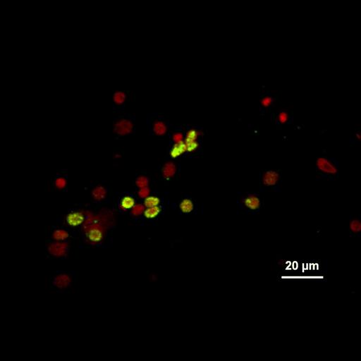
Aggregates of maize mosaic virus (MMV, Rhabdoviridae) in cells from hemolymph of the insect vector Peregrinus maidis (planthopper, Hemiptera, Delphacidae). Antibodies to MMV identify aggregates (green/yellow fluorescence) that accumulate mainly around the nuclei in hemocytes. Methods: Hemolymph smears were fixed in paraformaldehyde, labeled with primary antibodies to MMV, and with secondary antibodies: Alexa Fluor-488 (green), and later stained with the nuclear stain propidium iodide (red). Smears were examined with confocal laser- scanning microscope (Leica TCS SP). Associated references: Ammar, E.-D. and S. A. Hogenhout, S.A. (2008). A neurotropic route for Maize mosaic virus (Rhabdoviridae) in its planthopper vector Peregrinus maidis. Virus Research 131: 77-85. Ammar, E.-D. and Nault, L.R. (1985). Assembly and accumulation sites of Maize mosaic virus in its planthopper vector. Intervirology 24: 33-41.

Biological Sources
NCBI Organism Classification
Maize mosaic virus
Peregrinus maidis
Cell Type
hemocyte (sensu Nematoda and Protostomia)
Cellular Component
nucleus
host cell perinuclear region of cytoplasm
Biological Context
Biological Process
viral transmission by vector
Attribution
Names
El-Desouky Ammar
Saskia A. Hogenhout
L. R. Nault
Citation
Digital Object Identifier (DOI)
doi:10.7295/W9CIL25365
Archival Resource Key (ARK)
ark:/b7295/w9cil25365
Grouping This image is part of a group.
Imaging
Image Type
recorded image
Image Mode
confocal microscopy
Parameters Imaged
fluorescence emission
Source of Contrast
differences in adsorption or binding of stain
distribution of epitope
Visualization Methods
primary antibody plus labeled secondary antibody
Alexa Fluor 488
propidium iodide
Processing History
recorded image
Data Qualifiers
raw, unprocessed data
Sample Preparation
Methods
formaldehyde fixed tissue
permeabilized tissue
Dimensions
Spatial Axis Image Size Pixel Size
X 831px 0.21µm
Y 525px 0.21µm