Alternate header for print version


Licensing
Attribution Non-Commercial Share Alike:This image is licensed under a Creative Commons Attribution, Non-Commercial Share Alike License. View License Deed | View Legal Code
tweet  
*CIL – Cell Image Library accession number. Please use this to reference an image.

CIL:24805*  Cite 
Description

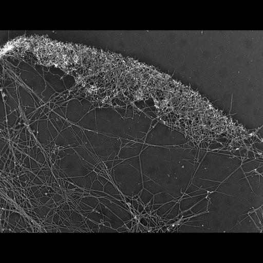
Differential response of lamellipodial actin network to latrunculin a (LA). Electron micrograph of a Xenopus fibroblast lamellipodium treated with LA (0.25 μM for 10 min) reveals actin depletion from the lamellipodial rear. A similar image of an LA treated keratocyte is available as CIL 24804. Image corresponds to Figure 7e from J Cell Biol. 1999 May 31;145(5):1009-26.

Technical Details

Procedures for detergent extraction, immunostaining, S1 decoration, light, and EM were described previously (Svitkina et al., 1995, 1996, 1997;Verkhovsky et al., 1995; Svitkina and Borisy, 1998).

Biological Sources
NCBI Organism Classification
Xenopus laevis
Cell Type
fibroblast
Cellular Component
actin cytoskeleton
lamellipodium
Biological Context
Biological Process
branching of actin filaments
latrunculin a treatment
actin filament-based process
Attribution
Names
Tatyana M. Svitkina
Gary G. Borisy
Published
J Cell Biol. 1999 May 31;145(5):1009-26.
Pubmed
10352018
Citation
Digital Object Identifier (DOI)
doi:10.7295/W9CIL24805
Archival Resource Key (ARK)
ark:/b7295/w9cil24805
Grouping This image is part of a group.
Imaging
Image Type
recorded image
Image Mode
transmission electron microscopy (TEM)
Parameters Imaged
optical path length gradient
Source of Contrast
boundaries between regions with different refractive index
Visualization Methods
shadowing and plating
platinum replica
Processing History
unprocessed raw data
Sample Preparation
Methods
permeabilized tissue
glutaraldehyde fixed tissue
phalloidin
Relation To Intact Cell
dispersed cells in vitro
Dimensions
Spatial Axis Image Size Pixel Size
X 2442px ——
Y 1968px ——