Licensing

Attribution_nc_sa
Attribution Non-Commercial Share Alike:This image is licensed under a Creative Commons Attribution, Non-Commercial Share Alike License. View License Deed | View Legal Code
Comments: Powered by Pnyxe

Description

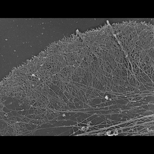
Structural differentiation of actin network in lamellipodium. Electron micrograph of Xenopus fibroblasts after regular extraction in the presence of polyethelene glycol (PEG) and phalloidin. While the actin network in the front zone of these cels remains dense, the actin network at the lamellipodial rear disassembled in the course of unprotected extraction (CIL 24800). Image corresponds to Figure 6c from J Cell Biol. 1999 May 31;145(5):1009-26.

Technical Details

Procedures for detergent extraction, immunostaining, S1 decoration, light, and EM were described previously (Svitkina et al., 1995, 1996, 1997;Verkhovsky et al., 1995; Svitkina and Borisy, 1998).

Biological Sources

NCBI Organism Classification
Xenopus laevis
(African clawed frog)
Cell Type
fibroblast
Cellular Component
lamellipodium
actin cytoskeleton
filopodium

Biological Context

Biological Process
cellular localization
actin filament organization
Molecular Function
structural constituent of cytoskeleton

Attribution

Names
Tatyana M. Svitkina
Gary G. Borisy
Published
J Cell Biol. 1999 May 31;145(5):1009-26.
Pubmed
10352018

Grouping

This image is part of a group.

Imaging

Image Type
recorded image
Imaging Mode
transmission electron microscopy (TEM)
Parameters Imaged
optical path length gradient
Source of Contrast
differences in deposition of metal shadow
Visualization Methods
shadowing and plating
platinum replica
Processing History
unprocessed raw data

Sample Preparation

Methods
permeabilized tissue
glutaraldehyde fixed tissue
phalloidin
polyethelene glycol
Relation To Intact Cell
dispersed cells in vitro

Dimensions

Spatial Axis Image Size Pixel Size
X 4380px ——
Y 3360px ——
*CIL – Cell Image Library accession number. Please use this to reference an image.