Licensing

Attribution_nc_sa
Attribution Non-Commercial Share Alike:This image is licensed under a Creative Commons Attribution, Non-Commercial Share Alike License. View License Deed | View Legal Code
Comments: Powered by Pnyxe

Description

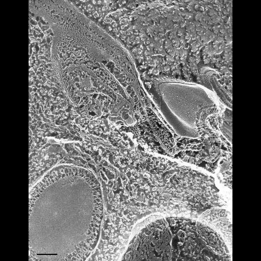
Quick-freeze deep-etch rotary shadowed replica of a parasomal sac from which a clathrin-coated preendosomal vesicle has pinched off. The typical clathrin cage structure is preserved. Fractured trichocyst at the lower left. TEM taken on 6/7/88 by C. Schroeder with Zeiss 10A operating at 80kV. Neg. 31,500X. Bar = 0.2µm. Adapted with permission from the J. Cell Sci. 101:449-461, 1992. A print of the negative was scanned and processed in Photoshop. This image is best used for qualitative analysis. A high resolution image (CIL:12633) is available for quantitative analysis. Additional information available at (http://www5.pbrc.hawaii.edu/allen/).

Biological Sources

NCBI Organism Classification
Paramecium multimicronucleatum
Cell Type
cell by organism
eukaryotic cell
Eukaryotic Protist
Ciliated Protist
Cellular Component
early endosome
clathrin coat
trichocyst

Attribution

Names
Richard Allen (University of Hawaii)
C. Schroeder
Published
J. Cell Sci. 101:449-461, 1992
Link
http://www5.pbrc.hawaii.edu/allen/
Pubmed
1629255

Grouping

This image is part of a group.
Data Qualifiers
processed data

Dimensions

Spatial Axis Image Size Pixel Size
X 2099px ——
Y 2686px ——
*CIL – Cell Image Library accession number. Please use this to reference an image.