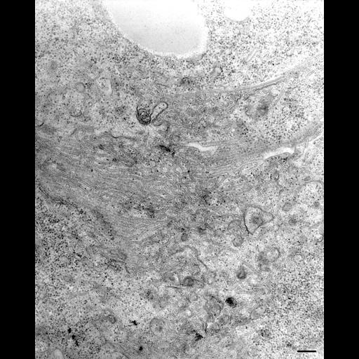
To determine what happens to the CV during systole we observed the CV in a living cell and then fixed the cell as soon as the CV had opened to the outside. It was apparent that the CV membrane does not flatten into an empty sac but the CV membrane undergoes rapid tubule formation and collapses as a continuous mat of tubules that maintains its connection to the microtubular ribbons. These tubules are a uniform 40nm in diameter. TEM taken on 6/9/96 by R. Allen with Zeiss 10A operating at 80kV. Neg. 19,800X. Bar = 0.2µm. Adapted with permission from the J. Exp. Biol. 200:1737-1744, 1997. The negative was printed to paper and the image was scanned to Photoshop. This digitized image is available for qualitative analysis. An unprocessed, high resolution version of this image (CIL:13113) is in the library and available for quantitative analysis. Standard glutaraldehyde fixation followed by osmium tetroxide, dehydrated in alcohol and embedded in an epoxy resin. Microtome sections prepared at approximately 75nm thickness. Additional information available at (http://www5.pbrc.hawaii.edu/allen/).
| Spatial Axis | Image Size | Pixel Size |
|---|---|---|
| X | 2051px | —— |
| Y | 2544px | —— |