Licensing
Description
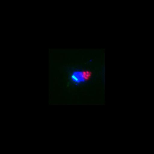
3HA-Cdc14 (red) localization in metaphase. The protein is sequestered in the nucleolus. Metaphase is determined by spindle morphology (tubulin, green) and nuclear morphology (DAPI, blue). Image is Fig 3D, left panels, in J Cell Biol. (2011) 192: 599-614. Images in Fig 3 include CIL# 13862, 13863, 13864, 13865, 13866, 13867.
Technical Details
Cells (MATa cdc14::3HA-CDC14), grown in rich media with glucose, were fixed for 15 min in 3.7% formaldehyde and 0.1 M potassium phosphate buffer, pH 6.4. Cells were then washed twice with 0.1 M potassium phosphate buffer, pH 6.4, and resuspended in 1.2 M sorbitol in 0.12 M K2HPO4/0.033 M citric acid, pH 5.9. Fixed cells were digested with 0.1 mg/ml zymolyase-100T (US Biological) and 1/10 volume of glusulase (PerkinElmer) at 30C for 15 min, washed once, and resuspended in 1.2 M sorbitol in 0.12 M K2HPO4/0.033 M citric acid, pH 5.9. Primary antibodies were anti-HA monoclonal antibody (HA.11; 1:500) and anti-tubulin (Abcam; 1:500). Secondary antibodies were: anti-mouse Cy3 (for HA) and anti-rat FITC (for tubulin). Cells were resuspended in DAPI (1 mg/ml). Imaging was performed at 25C using a Leica DM6000 microscope equipped with a 100x/1.40 NA oil immersion objective lens, A4, L5, and TX2 filters, and a digital CCD camera (DFC350, Leica). Pictures were processed with LAS AF (leica) and ImageJ software.
Biological Sources
- NCBI Organism Classification
- Saccharomyces cerevisiae
- (Baker's Yeast, Brewer's Yeast)
- Cell Line
- W303
- Cellular Component
- spindle pole body
- nucleus
- microtubule
- spindle pole body
- tubulin complex
- nucleolus
Biological Context
- Biological Process
- cell cycle
- protein dephosphorylation
- regulation of exit from mitosis
- mitotic sister chromatid segregation
- microtubule-based process
- Molecular Function
- protein tyrosine/serine/threonine phosphatase activity
- GTP binding
- GTPase activity
- nucleotide binding
- structural constituent of cytoskeleton
Attribution
- Names
- Mauricio Valerio-Santiago
- Fernando Monje-Casas
- Published
- J Cell Biol. (2011) 192: 599-614.
- Link
- http://jcb-dataviewer.rupress.org/jcb/...
- Pubmed
- 21321099
Grouping
This image is part of a group.Imaging
- Image Type
- recorded image
- Imaging Mode
- widefield illumination
- fluorescence microscopy
- Parameters Imaged
- fluorescence emission
- Source of Contrast
- distribution of a specific protein
- compartmentalization of stain or label
- Visualization Methods
- HA peptide tag
- 4',6-diamidino-2-phenylindole (DAPI)
- primary antibody plus labeled secondary antibody
- Cy3
- Fluorescein (FITC)
- Processing History
- unprocessed raw data
- Data Qualifiers
- raw, unprocessed data
- suitable for spatial measurements
Sample Preparation
- Methods
- formaldehyde fixed tissue
- Relation To Intact Cell
- whole mounted tissue
Dimensions
| Spatial Axis | Image Size | Pixel Size |
|---|---|---|
| X | 187px | .0642μm |
| Y | 187px | .0642μm |