Alternate header for print version


Licensing
Attribution Non-Commercial Share Alike:This image is licensed under a Creative Commons Attribution, Non-Commercial Share Alike License. View License Deed | View Legal Code
tweet  
*CIL – Cell Image Library accession number. Please use this to reference an image.

CIL:13701*  Cite 
Description

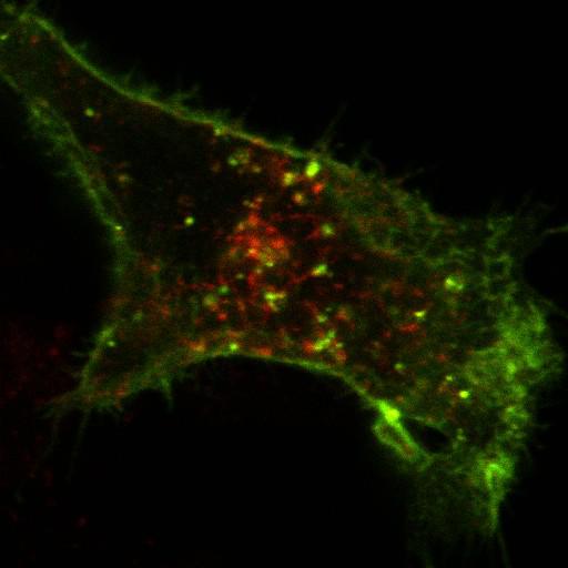
This is one of a group of four images in Figure S5 in Apaja et al., JCB 2010, that support the conclusion that the WT vasopressin type2 receptor (V2R) and the V2R W164S mutant (that causes nephrogenic diabetes insipidus) have different post-endocytic targeting. The sorting of endocytosed myc-tagged WT and W164S V2R was determined by indirect immunolabeling of internalized receptors in transiently transfected COS7 cells. The WT and temperature (26 degrees C) rescued mutant receptors were labelled with anti-myc Ab in live cells (1h) and then chased in Ab-free medium (1h, 37 degrees C) before indirect immunolabeling. Endosomes were labeled with 10 µg/ml Alexa Fluor 594–transferrin uptake for 1 h at 37°C. Fluorescence micrographs were obtained by a microscope (LSM510 or LSM710; Carl Zeiss, Inc.) equipped with a Plan-Apochromat 63×/NA 1.4 objective in multitrack mode. This single optical section shows that the internalized WT V2R is found in endosomes. Companion images show that the mutant V2R, however, co-localizes with lysosomes, but not endosomes.

Biological Sources
NCBI Organism Classification
Chlorocebus aethiops
Cell Line
COS-7
Cellular Component
plasma membrane
early endosome
Biological Context
Biological Process
G-protein coupled receptor internalization
Attribution
Names
Pirjo M. Apaja
Haijin Xu
Gergely L. Lukacs
Published
JCB 2010, 191:553-570
Pubmed
20974815
Citation
Digital Object Identifier (DOI)
doi:10.7295/W9CIL13701
Archival Resource Key (ARK)
ark:/b7295/w9cil13701
Grouping This image is part of a group.
Imaging
Image Type
recorded image
Image Mode
confocal microscopy
Parameters Imaged
fluorescence emission
Source of Contrast
distribution of a specific protein
Visualization Methods
Alexa Fluor 488
Alexa Fluor 594
Data Qualifiers
raw, unprocessed data
Sample Preparation
Methods
crosslinking-fixative fixed tissue
detergent permeabilized
Relation To Intact Cell
dispersed cells in vitro
Dimensions
Spatial Axis Image Size Pixel Size
X 512px 62nm
Y 512px 62nm