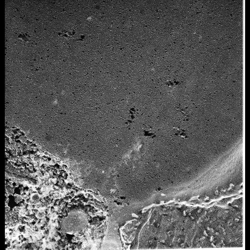
High resolution image of quick-freeze deep-etch of the P-face of the contractile vacuole. This face has many IMPs that are presumably transmembrane proteins many of which have been pulled free of the leaflet. There are also pits in this leaflet which represent the IMPs that remain with the opposite leaflet. Although we have found no mechano-sensitive or voltage-gated channels in the CVC (Sugino et al., J. Exp. Biol. 208:3957-3969, 2005) it is clear that proton pumps are present in the decorated tubules and that protons are exchanged for other ions such as calcium, potassium and sodium as these ions can accumulate in the CV against a concentration gradient from the cytosol. Some of the IMPs may function as ion exchange channels. What caused the large holes is not known. The texture of the luminal contents resembles the CV content and indicates the membrane is indeed that of the contractile vacuole. TEM taken on 5/27/92 by R. Allen with Zeiss 10A operating at 80kV. Neg. 31,500X. The raw film was scanned with an Epson Perfection V750 Pro. This image is best used for quantitative analysis. Additional information available at (http://www5.pbrc.hawaii.edu/allen/).
| Spatial Axis | Image Size | Pixel Size |
|---|---|---|
| X | 3740px | 0.63nm |
| Y | 3890px | 0.63nm |