Alternate header for print version


Licensing
This image is copyright protected. Any public or private use of this image is subject to prevailing copyright laws. Please contact the content provider of this image for permission requests.
tweet  
*CIL – Cell Image Library accession number. Please use this to reference an image.

CIL:12631*  Cite 
Description

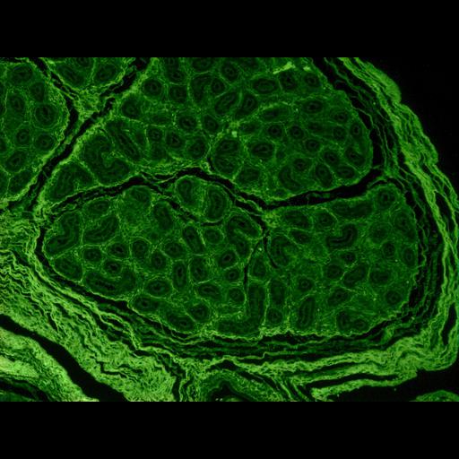
This cross section shows a human fetal testis. Numerous seminiferous tubules appear within a lobe of the testis. It has not been treated with an antibody but is autofluorescent and therefore appears bright green. The image was made from a commercially fixed and section slide that was stained with hematoxylin and eosin and imaged with a Nikon Eclipse inverted microscope

Biological Sources
NCBI Organism Classification
Homo sapiens
Biological Context
Human Development Anatomy
testis
seminiferous_tubules
Attribution
Names
Prue Talbot
Citation
Digital Object Identifier (DOI)
doi:10.7295/W9CIL12631
Archival Resource Key (ARK)
ark:/b7295/w9cil12631
Imaging
Image Type
recorded image
Image Mode
fluorescence microscopy
Parameters Imaged
fluorescence emission
Source of Contrast
non-specific protein affinity
Visualization Methods
hematoxylin
eosin
Processing History
unprocessed raw data
Data Qualifiers
raw, unprocessed data
Sample Preparation
Methods
chemically fixed tissue
Relation To Intact Cell
sectioned tissue
Dimensions
Spatial Axis Image Size Pixel Size
X 1600px ——
Y 1200px ——