Alternate header for print version


Licensing
Public Domain: This image is in the public domain and thus free of any copyright restrictions. However, as is the norm in scientific publishing and as a matter of courtesy, any user should credit the content provider for any public or private use of this image whenever possible. Learn more
tweet  
*CIL – Cell Image Library accession number. Please use this to reference an image.

CIL:12629*  Cite 
Description

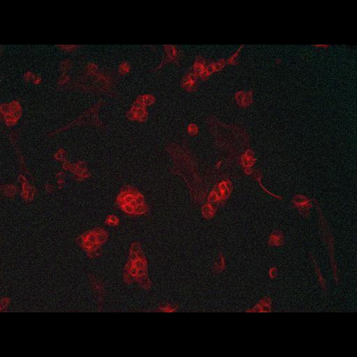
Thick microtubule bundles are organized around the periphery of each stem cell in human embryonic stem cell colonies. Human embryonic fibroblasts were grown on a layer of mouse embryonic fibroblasts which provide nutrients and help maintain pluripotency of the colonies. The sample has been labeled with an antibody to tubulin using standard protocols for immunolabeling.

Technical Details

Images were deconvolved using Nikon Elements software.

Biological Sources
NCBI Organism Classification
Homo sapiens
Cell Type
embryonic stem cell
Cellular Component
microtubule
Attribution
Names
Sabrina Lin
OTHER
UCR Stem Cell Center
Citation
Digital Object Identifier (DOI)
doi:10.7295/W9CIL12629
Archival Resource Key (ARK)
ark:/b7295/w9cil12629
Imaging
Image Type
recorded image
Image Mode
fluorescence microscopy
Parameters Imaged
fluorescence emission
Source of Contrast
distribution of epitope
Visualization Methods
primary antibody plus labeled secondary antibody
Processing History
deconvolution
Sample Preparation
Methods
chemically fixed tissue
Relation To Intact Cell
dispersed cells in vitro
Dimensions
Spatial Axis Image Size Pixel Size
X 1600px ——
Y 1200px ——