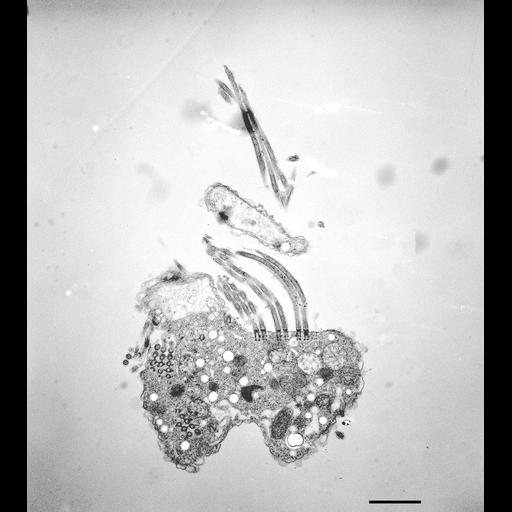
Halteria grandinella is a fresh water oligotrich ciliate. It lacks long rows of somatic cilia called kineties but has a few somatic ciliary bristles as well as a well developed oral and adoral or perioral ciliature. This section is a part of a serial series and the sections are thicker than normal so the EMs will appear less sharp when enlarged. For a more complete study of this cell refer to Grain, Protistologica 8:179-197, 1972. This figure is of a section near the cell’s left adoral surface where the contractile vacuole is located and where some of the oral ciliature are exposed in longitudinal section. A surface indentation may be near the CV pore. One membranelle of the perioral ciliature is sectioned through its basal bodies. The cytosol contains condensed as well as expanded mitochondria. The uniformly round and electron transparent structures seen here appear filled with dense material under different fixation and staining conditions. These may be pigment granules. TEM taken on 3/12/71 by R. Allen with Hitachi HU11A operating at 75kV. Neg. 3,800X. Bar = 2µm. Standard glutaraldehyde fixation followed by osmium tetroxide, dehydrated in alcohol and embedded in an epoxy resin. Thicker microtome sections prepared for a serial series. Additional information available at (http://www5.pbrc.hawaii.edu/allen/).
| Spatial Axis | Image Size | Pixel Size |
|---|---|---|
| X | 4288px | —— |
| Y | 4800px | —— |